中国畜牧兽医 ›› 2026, Vol. 53 ›› Issue (1): 427-438.doi: 10.16431/j.cnki.1671-7236.2026.01.038

• 基础兽医 • 上一篇    下一篇

蛋鸡肠黏膜损伤后抵御再次损伤的可能机制

齐思超1,2(), 于浩霖1, 张灵芝1, 华裕平1, 陈蕾晓1, 王华南1, 李剑1()   

  1. 1.浙江大学动物科学学院,农业农村部动物病毒学重点实验室,杭州 310058
    2.浙江大学海南研究院,三亚 572025
  • 收稿日期:2025-04-02 出版日期:2026-01-05 发布日期:2025-12-26
  • 通讯作者: 李剑 E-mail:scqi@zju.edu.cn;lijiannp@zju.edu.cn
  • 作者简介:齐思超,E-mail:scqi@zju.edu.cn
  • 基金资助:
    浙江省“三农九方”科技协作计划项目(2024SNJF046);国家自然科学基金面上项目(31972630)

Potential Mechanisms of Intestinal Mucosal Defense Against Secondary Injury in Laying Hens

QI Sichao1,2(), YU Haolin1, ZHANG Lingzhi1, HUA Yuping1, CHEN Leixiao1, WANG Huanan1, LI Jian1()   

  1. 1.MOA Key Laboratory of Animal Virology,College of Animal Sciences,Zhejiang University,Hangzhou 310058,China
    2.Hainan Institute of Zhejiang University,Sanya 572025,China
  • Received:2025-04-02 Online:2026-01-05 Published:2025-12-26
  • Contact: LI Jian E-mail:scqi@zju.edu.cn;lijiannp@zju.edu.cn

摘要:

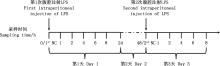
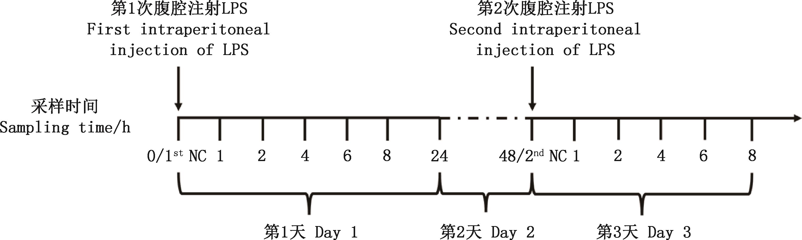
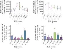
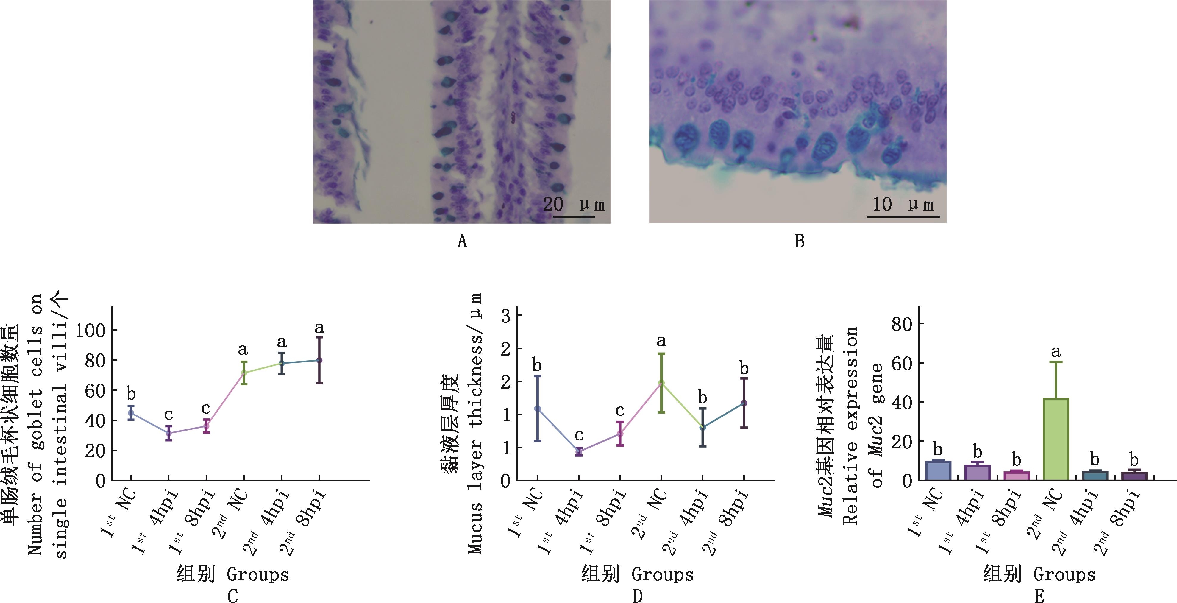
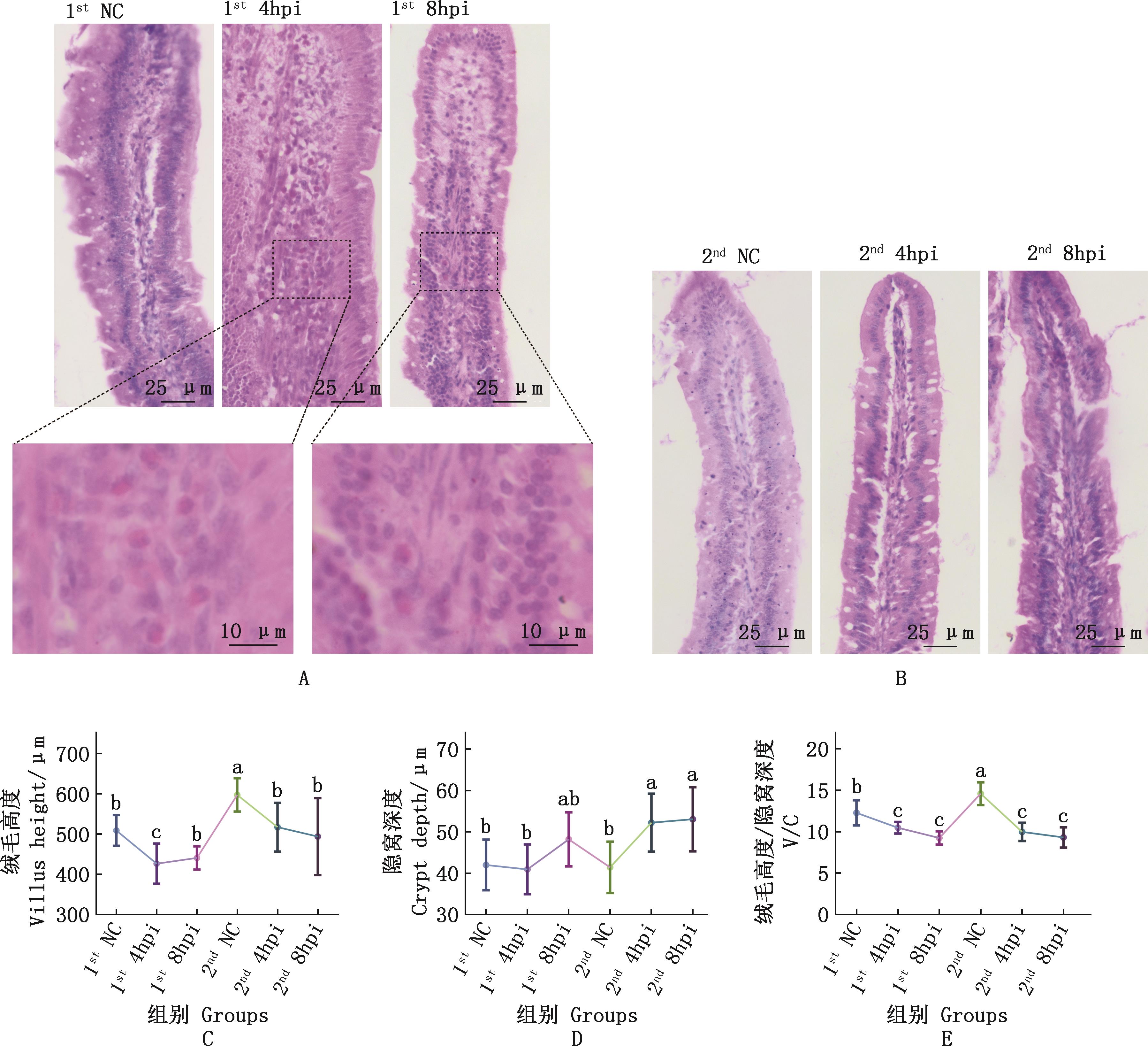
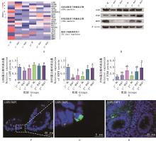
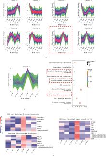
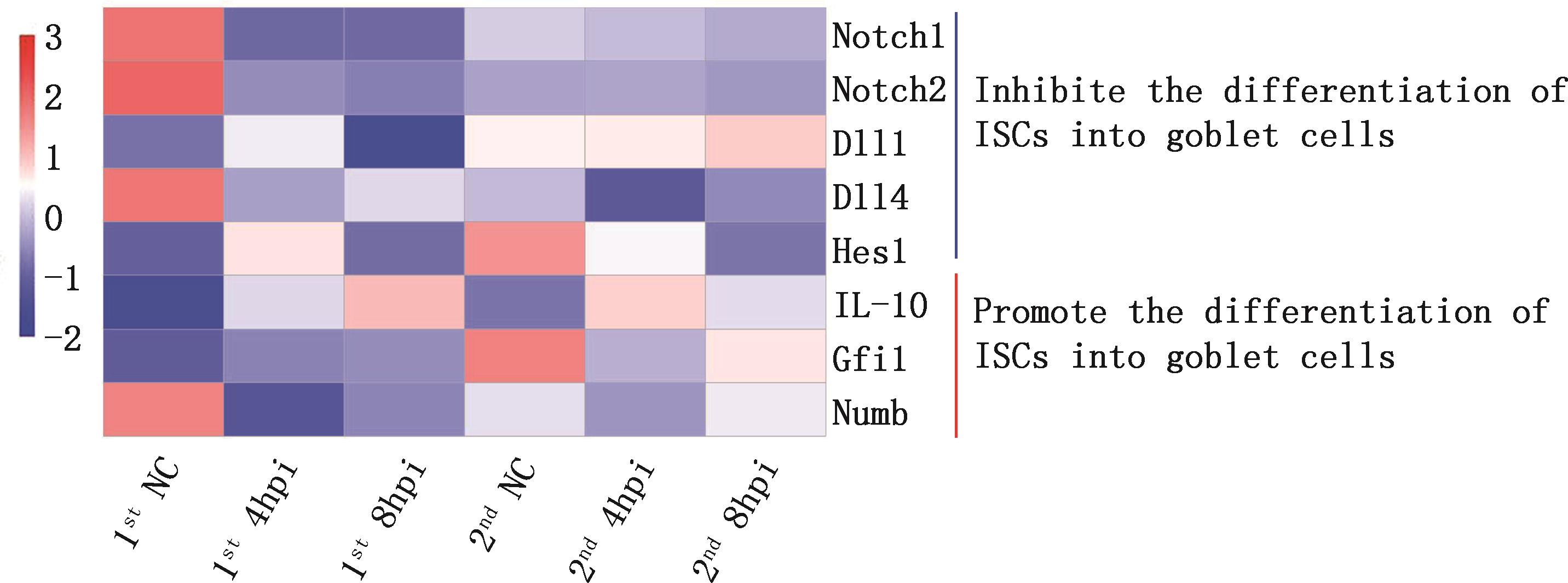
目的 研究蛋鸡肠黏膜在经历损伤后对再次损伤的抵抗力是否增强,并探究其可能机制,为探索促进蛋鸡肠黏膜损伤修复的手段提供新视角。 方法 选取78羽7日龄海兰白雏鸡,分为13个处理组(每组6羽),随机选取6羽雏鸡腹腔注射PBS作为阴性对照组,记为1st NC组;另外72羽雏鸡采用腹腔注射脂多糖(LPS,10 mg/kg BW)诱导肠黏膜损伤,在注射后1、2、4、6、8、24 h分别随机选取6羽雏鸡处死并采样,记为1st Xhpi(hours post injection)组(X代表注射后的采样时间)。在注射后48 h时,从注射LPS的雏鸡中随机选取6羽雏鸡腹腔注射PBS,作为第二阴性对照组,记为作2nd NC组(视同1st 48hpi组),剩余雏鸡第2次注射同剂量LPS。在第2次注射LPS后1、2、4、6、8 h分别随机选取6羽雏鸡处死并采样,分别记为2nd Xhpi组。所有组雏鸡采集血液、十二指肠组织并分离肠隐窝,分析其屏障功能、肠道干细胞(intestinal stem cells,ISCs)活性及隐窝微环境的变化。 结果 在LPS诱导损伤后,1st 4hpi组肠黏膜出现炎性细胞浸润、绒毛高度/隐窝深度比值下降、渗透性增加及黏液层厚度降低等损伤现象。注射后48 h,2nd NC组损伤程度减轻,屏障功能增强,表现为紧密连接Ocln基因表达量、杯状细胞密度、黏液层厚度及黏蛋白Muc2基因表达量均高于1st NC组。此时,进行第2次LPS诱导损伤,与2nd NC组相比,2nd 4hpi组肠黏膜的炎性细胞浸润程度与渗透性均未显著升高,但Claudin-1基因与补体蛋白C5基因表达量显著上调(P<0.05)。ISCs分析结果显示,1st 4hpi组活跃态ISCs(active intestinal stem cells,aISCs)活性下降,之后逐渐恢复,在损伤后48 h恢复正常;2nd 4hpi组休眠态ISCs(reserve intestinal stem cells,rISCs)可快速激活并维持较高活性。肠隐窝分析结果显示,在2次损伤过程中,肠隐窝Notch信号通路均受抑制。 结论 在经历损伤修复后,肠黏膜屏障功能增强,肠隐窝局部Notch信号通路受到抑制,第2次损伤可快速激活rISCs,并促进ISCs向分泌型上皮细胞分化。

关键词: 蛋鸡; 肠黏膜修复; 屏障功能; 肠道干细胞

Abstract:

Objective This study aimed to investigate whether the intestinal mucosa of laying hens developed enhanced resistance to secondary injury after initial injury and to explore the underlying mechanisms, thereby promoting new perspectives for developing approaches to repair intestinal mucosal damage in laying hens. Method Seventy-eight 7-day-old Hyline White chicks were divided into 13 treatment groups (6 chicks per group). Six chicks were radomly selected and received an intraperitoneal injection of PBS as the first negative control group (1st NC). The remaining 72 chicks were subjected to intestinal mucosal injury via intraperitoneal injection of lipopolysaccharide (LPS, 10 mg/kg BW). Six chicks were radomly selected, euthanized and sampled at 1, 2, 4, 6, 8 and 24 hours post-injection (hpi), designated as 1st Xhpi groups (X represents the time of sampling after injection). At 48 hours post-injection, six chicks from the LPS-injected group were radomly selected and injected with PBS as the second negative control group (2nd NC, equivalent to the 1st 48hpi group), the remaining chicks received a second injection of the same dose of LPS. Six chicks were radomly selected, euthanized and sampled at 1, 2, 4, 6 and 8 hours post the second LPS challenge, designated as 2nd Xhpi groups. Blood, duodenal tissues were collected, and intestinal crypts were isolated for analysis of barrier function, intestinal stem cells (ISCs) activity, and crypt niche changes. Result Following the first LPS challenge, inflammatory cell infiltration, decreased villus height/crypt depth, increased permeability, and decreased mucus layer thickness were observed in the intestinal mucosa of the 1st 4hpi group. At 48 hours post injection, the degree of injury in the 2nd NC group was reduced, and the barrier function was enhanced, which was manifested as the expression level of the tight junction Ocln gene, the density of goblet cells, the thickness of the mucus layer, and the expression level of the mucin Muc2 gene were all higher than those in the 1st NC group. After the second LPS challenge, compared with 2nd NC group,in the 2nd 4hpi group, inflammatory cell infiltration and permeability did not significantly increase, but Claudin-1 and complement protein C5 gene levels were markedly upregulated (P<0.05). The ISCs analysis results showed that the activity of active intestinal stem cells (aISCs) in the 1st 4hpi group decreased and then gradually recovered, returning to normal at 48 hours post injury.The reserve ISCs (rISCs) in the 2nd 4hpi group could be rapidly activated and maintain high activity. The analysis results of intestinal crypts showed that the Notch signal pathway of intestinal crypts was inhibited in both injury processes. Conclusion Following initial injury and repair, the intestinal mucosa exhibited enhanced barrier function and suppressed Notch signaling pathway in crypts. Secondary injury triggered rapid activation of rISCs and promoted differentiation of ISCs into secretory epithelial cells.

Key words: laying hens; intestinal mucosal repair; intestinal barrier function; intestinal stem cells

中图分类号: