
中国畜牧兽医 ›› 2026, Vol. 53 ›› Issue (2): 659-670.doi: 10.16431/j.cnki.1671-7236.2026.02.014
张之盈1( ), 胡晓迪1, 彭苏1, 张兴月1, 佘沛宁1, 黄小洁2, 石达友1()
收稿日期:2025-07-15
出版日期:2026-02-05
发布日期:2026-01-27
通讯作者:
石达友
E-mail:2549215237@qq.com;shidayou@scau.edu.cn
作者简介:张之盈,E-mail: 2549215237@qq.com
基金资助:
ZHANG Zhiying1(), HU Xiaodi1, PENG Su1, ZHANG Xingyue1, SHE Peining1, HUANG Xiaojie2, SHI Dayou1()
Received:2025-07-15
Online:2026-02-05
Published:2026-01-27
Contact:
SHI Dayou
E-mail:2549215237@qq.com;shidayou@scau.edu.cn
摘要:
目的 探讨腹腔注射脂多糖(LPS)诱导麻黄鸡炎症模型的最佳剂量。 方法 选取120只1日龄(39.11 g±3.13 g)雄性麻黄鸡,随机分为4组,每组6个重复,每个重复5只鸡,试验期21 d。对照组(CON)麻黄鸡在15、17、19和21日龄时腹腔注射生理盐水,试验组(LL、LM和LH)分别腹腔注射0.25、0.5和1 mg/kg LPS。于15和21 d进行采血并屠宰,计算生长性能指标和免疫器官指数,观察空肠形态,测定血清生化指标、皮质酮含量、炎症因子水平、抗氧化指标及相关基因表达量。 结果 15 d时,与CON组相比,各LPS处理组麻黄鸡终末体重和平均日增重均无显著变化(P>0.05);LH组麻黄鸡空肠绒毛高度(VH)和绒隐比(V/C)均显著下降(P<0.05);LM和LH组麻黄鸡空肠闭锁蛋白(Occludin)和核因子-κB(NF-κB)表达均显著下调(P<0.05);LH组麻黄鸡血清皮质酮含量显著升高(P<0.05);LL、LM和LH组麻黄鸡血清甘油三酯(TG)含量均显著降低(P<0.05);LH组麻黄鸡血清细胞炎症因子白细胞介素-6(IL-6)含量显著升高(P<0.05);LH组麻黄鸡血清总超氧化物歧化酶(T-SOD)活性显著降低(P<0.05),丙二醛(MDA)含量显著升高(P<0.05)。21 d时,与CON组相比,LM和LH组麻黄鸡脾脏指数和胸腺指数均有所升高(P>0.05);LH组麻黄鸡空肠隐窝深度(CD)极显著升高(P<0.01),V/C显著降低(P<0.05);LH组麻黄鸡空肠闭合蛋白1(Claudin-1)表达极显著下调(P<0.01),IL-1β、肿瘤坏死因子-α(TNF-α)和NF-κB表达均极显著上调(P<0.01);各LPS组T-SOD活性均显著降低(P<0.05)。 结论 LPS注射可显著影响麻黄鸡免疫器官发育、肠道形态、炎症因子表达及抗氧化功能,成功诱导炎症应激。综合各项指标,1.0 mg/kg LPS为构建麻黄鸡炎症模型的最佳剂量。
中图分类号:
张之盈, 胡晓迪, 彭苏, 张兴月, 佘沛宁, 黄小洁, 石达友. LPS对麻黄鸡生长性能、血清生化及肠道健康的影响[J]. 中国畜牧兽医, 2026, 53(2): 659-670.
ZHANG Zhiying, HU Xiaodi, PENG Su, ZHANG Xingyue, SHE Peining, HUANG Xiaojie, SHI Dayou. Effects of LPS on the Growth Performance, Serum Biochemistry and Intestinal Health of Mahuang Chickens[J]. China Animal Husbandry & Veterinary Medicine, 2026, 53(2): 659-670.
表1
基础饲粮组成及营养水平(风干基础) (%)"
| 原料 Ingredients | 含量 Contents | 营养成分 Nutrient components② | 含量 Contents |
|---|---|---|---|
| 玉米 Corn | 54.50 | 代谢能 ME/(MJ/kg) | 12.22 |
| 豆粕 Soybeanmeal (CP, 43%) | 33.00 | 粗蛋白质 CP | 21.00 |
| 鱼粉 Fish meal | 1.00 | 钙 Calcium | 1.05 |
| 麦麸 Wheat bran | 5.50 | 钠 Sodium | 0.16 |
| 大豆油 Soy oil | 1.50 | 氯气 Chlorine | 0.16 |
| 菜籽粕 Rapeseed meal | 0.00 | 总磷 Total phosphorus | 0.50 |
| 石灰石粉 Limestone powder | 1.40 | 赖氨酸 Lysine | 1.20 |
| 磷酸氢钙 CaHPO4 | 1.10 | 甲硫氨酸 Methionine | 0.55 |
| 预混料 Premix① | 2.00 | 苏氨酸 Threonine | 0.19 |
| 总计 Total | 100.00 |
表2
实时荧光定量PCR引物序列"
基因 Genes | 引物序列 Primer sequences (5'→3') | |
|---|---|---|
| β-actin | F:GAGAAATTGTGCGTGACATCA | R:CCTGAACCTCTCATTGCCA |
| IL-1β | F:GAAGTGCTTCGTGCTGGAGT | R:ACTGGCATCTGCCCAGTTC |
| TNF-α | F:AATTTGCAGGCTGTTTCTGC | R:TATGAAGGTGGTGCAGATGG |
| Claudin-1 | F:GAGGATGACCAGGTCAAGAAG | R:TGCCCAGCCAATGAAGAG |
| ZO-1 | F:CCAAAGACAGCAGGAGGAGA | R:TGGCTAGTTTCTCTCGTGCA |
| Occludin | F:TCATCCTGCTCTGCCTCATCT | R:CATCCGCCACGTTCTTCAC |
| sIgA | F:TCCTTCCTCCCTTCTTCCATC | R:TTTGCCAATCCTCCACGG |
| NF-κB | F:CAGCCCATCTATGACAACCG | R:TCAGCCCAGAAACGAACCTC |
表3
LPS对麻黄鸡生长性能的影响 (g)"
| 项目 Items | 组别 Groups | P值 P-value | |||
|---|---|---|---|---|---|
| CON | LL | LM | LH | ||
| 体重 Body weight | |||||
| 0 d | 39.88±3.63 | 39.25±1.91 | 39.25±3.00 | 38.06±3.66 | 0.429 |
| 15 d | 236.00±16.63 | 222.00±14.91 | 220.00±13.99 | 230.17±18.39 | 0.309 |
| 21 d | 374.00±50.19 | 318.17±49.10 | 319.00±64.32 | 337.33±37.54 | 0.229 |
| 平均日增重 ADG | |||||
| 0~15 d | 11.61±0.94 | 12.21±0.77 | 11.65±0.31 | 12.23±0.45 | 0.523 |
| 15~21 d | 20.24±4.10 | 17.10±2.17 | 16.83±2.52 | 16.31±2.08 | 0.377 |
| 0~21 d | 15.04±0.73 | 14.43±0.53 | 13.93±0.70 | 14.17±0.68 | 0.275 |
表4
LPS对麻黄鸡免疫器官指数的影响 (%)"
| 项目 Items | 组别 Groups | P值 P-value | |||
|---|---|---|---|---|---|
| CON | LL | LM | LH | ||
| 15 d | |||||
| 脾脏指数 Spleen index | 1.24±0.23 | 1.64±0.45 | 1.46±0.39 | 1.37±0.39 | 0.331 |
| 胸腺指数 Thymus index | 4.10±0.23 | 4.28±0.45 | 4.04±0.46 | 4.44±0.85 | 0.576 |
| 法氏囊指数 Bursa of Fabricius index | 3.17±0.83 | 3.05±0.51 | 2.47±0.57 | 2.86±0.58 | 0.233 |
| 21 d | |||||
| 脾脏指数 Spleen index | 2.00±0.35 | 1.70±0.41 | 2.42±0.46 | 2.36±1.00 | 0.177 |
| 胸腺指数 Thymus index | 5.82±0.60a | 4.72±0.52b | 5.94±0.51a | 6.27±0.86a | 0.026 |
| 法氏囊指数 Bursa of Fabricius index | 3.94±1.63 | 4.01±1.01 | 2.92±0.77 | 3.12±0.57 | 0.215 |
表5
LPS对麻黄鸡空肠绒毛的影响"
| 项目Items | 组别Groups | P值 P-value | |||
|---|---|---|---|---|---|
| CON | LL | LM | LH | ||
| 15 d | |||||
| 绒毛高度VH/μm | 1 052.58±34.73Aa | 978.75±24.31Aa | 1 106.33±229.61Aa | 707.25±92.13Bb | 0.003 |
| 隐窝深度CD/μm | 151.49±2.92 | 153.83±9.48 | 163.45±9.12 | 156.07±6.66 | 0.184 |
| 绒毛高度/隐窝深度V/C | 6.95±0.18a | 6.38±0.44a | 6.81±1.64a | 4.56±0.77b | 0.012 |
| 21 d | |||||
| 绒毛高度VH/μm | 985.31±42.36 | 995.17±75.18 | 1 038.94±70.63 | 935.22±28.73 | 0.142 |
| 隐窝深度CD/μm | 126.32±6.33Cc | 134.82±4.21Cc | 157.36±19.46Bb | 176.78±4.31Aa | <0.001 |
| 绒毛高度/隐窝深度V/C | 7.81±0.37Aa | 7.40±0.74Aa | 6.72±1.26Aa | 5.29±0.05Bb | 0.003 |
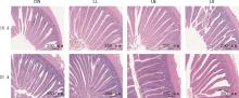
图1
LPS对麻黄鸡空肠绒毛的影响(200×)"

表6
LPS对麻黄鸡空肠免疫和屏障基因表达的影响"
| 项目 Items | 组别 Groups | P值 P-value | |||
|---|---|---|---|---|---|
| CON | LL | LM | LH | ||
| 15 d | |||||
| Claudin-1 | 0.97±0.50 | 0.93±0.63 | 0.48±0.47 | 0.40±0.18 | 0.106 |
| Occludin | 1.05±0.56Aa | 1.11±0.41Aa | 0.36±0.23Bb | 0.50±0.25Bb | 0.005 |
| ZO-1 | 1.00±0.32 | 0.96±0.32 | 0.63±0.51 | 0.70±0.45 | 0.310 |
| IL-1β | 1.31±0.56 | 0.65±0.16 | 1.02±0.69 | 1.03±0.28 | 0.606 |
| NF-κB | 1.14±0.4Aa | 0.76±0.24ABab | 0.48±0.4Bb | 0.42±0.24Bb | 0.005 |
| TNF-α | 0.96±0.27 | 0.65±0.35 | 0.96±0.69 | 0.68±0.40 | 0.499 |
| sIgA | 1.55±0.86 | 0.36±0.07 | 1.13±0.53 | 1.01±0.28 | 0.135 |
| 21 d | |||||
| Claudin-1 | 1.15±0.52Aa | 0.25±0.12Bb | 1.16±0.53Aa | 0.47±0.30Bb | 0.001 |
| Occludin | 1.01±0.16ab | 0.57±0.19b | 1.24±0.23a | 1.20±0.25a | 0.021 |
| ZO-1 | 1.03±0.31 | 0.72±0.31 | 0.96±0.08 | 0.78±0.20 | 0.104 |
| IL-1β | 1.31±0.58Bb | 1.55±0.47Bb | 1.47±0.66Bb | 6.81±3.48Aa | <0.001 |
| NF-κB | 1.03±0.30Bb | 0.70±0.20Bb | 0.99±0.09Bb | 1.86±0.56Aa | <0.001 |
| TNF-α | 0.93±0.40Bb | 0.68±0.18Bb | 0.56±0.06Bb | 2.15±0.99Aa | <0.001 |
| sIgA | 1.14±0.77ab | 0.39±0.18b | 1.34±0.42ab | 1.88±0.59a | 0.018 |
表7
LPS对麻黄鸡血清生化的影响"
| 项目 Items | 组别 Groups | P值 P-value | |||
|---|---|---|---|---|---|
| CON | LL | LM | LH | ||
| 15 d | |||||
| 尿素 UREA/(mmol/L) | 0.99±0.21 | 1.15±0.39 | 1.10±0.31 | 1.13±0.25 | 0.803 |
| 碱性磷酸酶 ALP/(U/L) | 11 792.00±3 904.79 | 10 501.96±4 581.57 | 12 384.38±4 520.21 | 8 035.85±3 174.86 | 0.491 |
| 天冬氨酸氨基转移酶 AST/(U/L) | 388.27±67.94 | 341.47±58.45 | 247.27±33.13 | 438.73±93.873 | 0.261 |
| 丙氨酸氨基转移酶 ALT/(U/L) | 4.03±1.63 | 4.40±1.58 | 3.42±0.94 | 4.65±1.57 | 0.503 |
| 总胆固醇 TC/(mmol/L) | 4.55±0.78 | 3.38±0.97 | 3.78±0.68 | 3.78±0.68 | 0.054 |
| 甘油三酯 TG/(mmol/L) | 1.41±0.29Aa | 0.71±0.29Bb | 0.80±0.41Bb | 0.56±0.20Bb | 0.001 |
| 血清白蛋白ALB Ⅱ/(g/L) | 11.78±1.99 | 7.78±3.71 | 10.40±2.40 | 10.45±2.44 | 0.110 |
| 总蛋白 TP-Ⅱ/(g/L) | 30.13±3.47 | 25.75±4.61 | 27.08±5.61 | 26.40±5.15 | 0.418 |
| 白蛋白 GLB/(g/L) | 18.17±1.47 | 16.17±2.48 | 16.83±3.25 | 15.83±2.64 | 0.418 |
| 21 d | |||||
| 尿素 UREA/(mmol/L) | 0.60±0.11 | 0.68±0.16 | 0.63±0.15 | 0.70±0.36 | 0.868 |
| 碱性磷酸酶 ALP/(U/L) | 3 435.60±657.14 | 7 003.47±1 910.05 | 6 466.52±1 461.62 | 3 413.35±1 266.70 | 0.190 |
| 天冬氨酸氨基转移酶 AST/(U/L) | 228.83±23.22 | 247.93±39.87 | 224.78±35.03 | 305.01±64.69 | 0.456 |
| 丙氨酸氨基转移酶 ALT/(U/L) | 3.28±1.79 | 3.92±1.14 | 3.70±0.81 | 4.72±1.59 | 0.361 |
| 总胆固醇 TC/(mmol/L) | 3.76±0.48 | 3.69±0.61 | 3.68±0.60 | 3.58±0.64 | 0.964 |
| 甘油三酯 TG/(mmol/L) | 0.32±0.07 | 0.34±0.07 | 0.36±0.03 | 0.34±0.07 | 0.693 |
| 血清白蛋白 ALB Ⅱ/(g/L) | 11.48±1.77 | 1.83±2.14 | 12.12±1.45 | 10.16±2.94 | 0.460 |
| 总蛋白 TP-Ⅱ/(g/L) | 31.57±5.16 | 33.92±5.82 | 35.33±8.98 | 28.87±8.99 | 0.474 |
| 白蛋白 GLB/(g/L) | 20.00±3.52 | 22.00±3.84 | 23.33±7.71 | 18.67±5.54 | 0.499 |
表8
LPS对麻黄鸡皮质酮的影响 (ng/mL)"
日龄 Age | 组别 Groups | P值 P-value | |||
|---|---|---|---|---|---|
| CON | LL | LM | LH | ||
| 15 d | 337.93±39.17b | 359.97±33.73ab | 352.55±37.87b | 388.88±21.95a | 0.039 |
| 21 d | 363.19±47.80 | 377.68±40.10 | 357.58±28.11 | 395.58±25.80 | 0.185 |
表9
LPS对麻黄鸡血清细胞因子的影响 (pg/mL)"
| 项目 Items | 组别 Groups | P值 | |||
|---|---|---|---|---|---|
| CON | LL | LM | LH | P-value | |
| 15 d | |||||
| 肿瘤坏死因子-α TNF-α | 0.14±0.00 | 0.13±0.01 | 0.14±0.01 | 0.14±0.00 | 0.252 |
| 白细胞介素-6 IL-6 | 1.39±0.05Bb | 1.39±0.08Bb | 1.43±0.07Bb | 1.59±0.14Aa | 0.008 |
| 白细胞介素-1β IL-1β | 1.20±0.02 | 1.19±0.03 | 1.19±0.02 | 1.21±0.02 | 0.503 |
| 21 d | |||||
| 肿瘤坏死因子-α TNF-α | 0.15±0.03 | 0.17±0.01 | 0.15±0.02 | 0.18±0.04 | 0.414 |
| 白细胞介素-6 IL-6 | 1.68±0.20 | 1.70±0.17 | 1.87±0.27 | 2.00±0.51 | 0.523 |
| 白细胞介素-1β IL-1β | 1.24±0.07 | 1.24±0.04 | 1.22±0.03 | 1.29±0.07 | 0.308 |
表10
LPS对麻黄鸡抗氧化的影响"
| 项目 Items | 组别 Groups | P值 P-value | |||
|---|---|---|---|---|---|
| CON | LL | LM | LH | ||
| 15 d | |||||
| 总超氧化物歧化酶 T-SOD/(U/mg prot) | 2.88±0.56a | 2.41±0.47b | 2.60±0.13ab | 2.44±0.21b | 0.060 |
| 总抗氧化能力 T-AOC/(U/mg prot) | 0.11±0.07b | 0.19±0.04a | 0.15±0.05ab | 0.20±0.04a | 0.016 |
| 丙二醛 MDA/(nmol/mg prot) | 0.05±0.01b | 0.09±0.03a | 0.08±0.03a | 0.09±0.03a | 0.028 |
| 过氧化氢酶 CAT/(U/mg prot) | 0.47±0.19 | 0.25±0.17 | 0.43±0.19 | 0.43±0.17 | 0.129 |
| 21 d | |||||
| 总超氧化物歧化酶 T-SOD/(U/mg prot) | 3.13±0.31Aa | 2.49±0.25Bb | 2.77±0.29Bb | 2.68±0.06Bb | 0.003 |
| 总抗氧化能力 T-AOC/(U/mg prot) | 0.33±0.09ab | 0.38±0.06a | 0.29±0.05b | 0.40±0.10a | 0.048 |
| 丙二醛 MDA/(nmol/mg prot) | 0.03±0.02 | 0.10±0.06 | 0.12±0.09 | 0.11±0.13 | 0.288 |
| 过氧化氢酶 CAT/(U/mg prot) | 1.19±0.13Aa | 0.89±0.16Bb | 0.88±0.21Bb | 1.07±0.03ABab | 0.003 |
| [1] | 张朝生, 陈志敏, 王少龙, 等. 白头翁口服液对脂多糖攻毒无特定病原体白来航鸡肠道炎症以及氧化应激的影响[J]. 动物营养学报, 2025, 37(3): 1771-1783. |
| ZHANG C S, CHEN Z M, WANG S L, et al. Effects of Baitouweng oral liquid on intestinal inflammation and oxidative stress of lipopolysaccharide challenge specific pathogen free White Leghorn chickens[J]. Chinese Journal of Animal Nutrition, 2025, 37(3): 1771-1783. (in Chinese) | |
| [2] | 高嘉豪, 乔彦杰, 连科迅, 等. 甘草酸对脂多糖应激黄羽肉仔鸡免疫性能的影响[J]. 中国畜牧兽医, 2022, 49(9): 3419-3427. |
| GAO J H, QIAO Y J, LIAN K X, et al. Effects of glycyrrhizic acid on immune performance of lipopolysaccharide-stressed Yellow feather broilers[J]. China Animal Husbandary & Veterinary Medicine, 2022, 49(9): 3419-3427. (in Chinese) | |
| [3] | JIN S, WANG H, GONG H, et al. Music intervention mitigates LPS-induced gut barrier disruption and immune stress in broilers via TLR4/NF-κB regulation[J]. Poultry Science, 2025, 104(7): 105189. |
| [4] | 陆瑞, 盛辉, 郭妍岩, 等. 脂多糖免疫应激研究进展[J]. 中国畜牧兽医, 2025, 52(2): 946-958. |
| LU R, SHENG H, GUO Y Y, et al. Research progress on immune stress of LPS[J]. China Animal Husbandary & Veterinary Medicine, 2025, 52(2): 946-958. (in Chinese) | |
| [5] | 陈乐乐, 陈博, 伍新珍, 等. 肉桂醛对热应激肉鸡生长性能、抗氧化能力和肠道健康的影响[J]. 动物营养学报, 2024, 36(9): 5642-5655. |
| CHEN L L, CHEN B, WU X Z, et al. Effects of cinnamaldehyde on growth performance, antioxidant capacity and intestinal health of broilers under heat stress[J]. Chinese Journal of Animal Nutrition, 2024, 36(9): 5642-5655. (in Chinese) | |
| [6] | 杜林, 杨萍瑞, 周翰林, 等. 三七提取物通过Il-6、CASP3和STAT3调节免疫应激肉鸡炎症反应[J]. 中国兽医学报, 2024, 44(8): 1755-1764. |
| DU L, YANG P R, ZHOU H L, et al. Panax notoginseng extracts regulate inflammatory response of immune-stressed broilers through IL-6,CASP3 and STAT3[J]. Chinese Journal of Veterinary Science, 2024, 44(8): 1755-1764. (in Chinese) | |
| [7] | 江皓天, 周思源, 王雨佳, 等. 异绿原酸对肉鸡生长性能、养分表观代谢率及脂多糖应激状态下抗氧化能力的影响[J]. 动物营养学报, 2024, 36(9): 5630-5641. |
| JIANG H T, ZHOU S Y, WANG Y J, et al. Effects of isochlorogenic acid on growth performance, nutrient apparent metabolic rates of broilers and antioxidant capacity of broilers under lipopolysaccharide stress[J]. Chinese Journal of Animal Nutrition, 2024, 36(9): 5630-5641. (in Chinese) | |
| [8] | 樊佳奇, 韩雯笑, 范慧敏, 等. 酵母肽对肉兔生长性能以及脂多糖应激肉兔血清细胞因子和肝脏抗氧化能力的影响[J]. 动物营养学报, 2024, 36(8): 5308-5318. |
| FAN J Q, HAN W X, FAN H M, et al. Effects of yeast peptides on growth performance of meat rabbits, and serum cytokines and liver antioxidant capacity of lipopolysaccharide-stressed meat rabbits[J]. Chinese Journal of Animal Nutrition, 2024, 36(8): 5308-5318. (in Chinese) | |
| [9] | LI Y, ZHANG H, CHEN Y P, et al. Bacillus amyloliquefaciens supplementation alleviates immunological stress and intestinal damage in lipopolysaccharide-challenged broilers[J]. Animal Feed Science and Technology, 2015, 208: 119-131. |
| [10] | 张柏林, 杨乾, 刘宁, 等. 饲粮添加L-谷氨酰胺对脂多糖刺激肉鸡血浆生化指标、免疫性能、肠道炎症因子表达及黏膜免疫的影响[J]. 动物营养学报, 2020, 32(6): 2611-2623. |
| ZHANG B L, YANG Q, LIU N, et al. Effects of dietary L-glutamine supplementation on plasma biochemical parameters, immune performance, intestinal inflammatory factors expression and mucosal immune of broilers challenged by lipopolysaccharide[J]. Chinese Journal of Animal Nutrition, 2020, 32(6): 2611-2623. (in Chinese) | |
| [11] | 廖莹, 李悦伊, 刘巍, 等. 不同品种广西黄羽肉鸡与AA肉鸡风味物质及口感的比较[J]. 饲料研究, 2024, 47(12): 105-110. |
| LIAO Y, LI Y Y, LIU W, et al, Comparison of flavor substances and taste of different varieties of Guangxi Yellow-feathered broilers and AA broilers[J]. Feed Research, 2024, 47(12): 105-110. (in Chinese) | |
| [12] | XIAO L, QI L, FU R, et al. A large-scale comparison of the meat quality characteristics of different chicken breeds in South China[J]. Poultry Science, 2024, 103(6): 103740. |
| [13] | 叶茂, 江伟烽, 许宇航, 等. 两个麻黄鸡纯系体尺性状和屠宰性能比较及相关性分析[J]. 中国家禽, 2023, 45(7): 107-111. |
| YE M, JIANG W F, XU Y H, et al. Comparison and correlation analysis of body size traitsand slaughter performance of two pure strains of Mahuang chickens[J]. China Poultry, 2023, 45(7): 107-111. (in Chinese) | |
| [14] | 冯焱, 张芬鹊, 薛智全, 等. 脂多糖和地塞米松对肉鸡生长性能、养分代谢、血清生化指标及肠道形态发育的影响[J]. 中国畜牧兽医, 2017, 44(3): 732-739. |
| FENG Y, ZHANG F Q, XUE Z Q, et al. Effects of glycyrrhizic acid on immune performance of lipopolysaccharide-stressed Yellow feather broilers[J]. China Animal Husbandary & Veterinary Medicine, 2017, 44(3): 732-739. (in Chinese) | |
| [15] | 刘杰. 不同TI表型肉鸡血液生化指标的检测及对LPS刺激反应性的研究[D].南京:南京农业大学, 2018. |
| LIU J. Studies on blood bioparameters correlated with TI duration and stress responses to LPS stimulation in different TI phenotype of broilers[D]. Nanjing: Nanjing Agricultural University, 2018. (in Chinese) | |
| [16] | SONG W, CHEN J, AI G, et al. Mechanisms of the effects of turpiniae folium extract on growth performance, immunity, antioxidant activity and intestinal barrier function in LPS-challenged broilers[J]. Poultry Science, 2025, 104(4): 104903. |
| [17] | ZHENG X C, WU Q J, SONG Z H, et al. Effects of oridonin on growth performance and oxidative stress in broilers challenged with lipopolysaccharide[J]. Poultry Science, 2016, 95(10): 2281-2289. |
| [18] | QU Q, LIU M, HU Y, et al. Modulatory effects of polyherbal mixture on the immuno-antioxidant capacity and intestinal health of chicks infected with Escherichia coli O78[J]. Poultry Science, 2025, 104(6): 105156. |
| [19] | 阳金金, 杨芷, 杨雨, 等. 甜菜碱对脂多糖刺激仔鹅生长性能、器官指数、血清生化指标及脾脏炎性因子表达的影响[J]. 动物营养学报, 2021, 33(4): 2044-2054. |
| YANG J J, YANG Z, YANG Y, et al. Effects of betaine on growth performance, organ indices, serum biochemical parameters and spleen inflammatory factor mRNA expression of geese challenged by lipopolysaccharide[J]. Chinese Journal of Animal Nutrition, 2021, 33(4): 2044-2054. (in Chinese) | |
| [20] | XIE Y, WEN M, ZHAO H, et al. Effect of zinc supplementation on growth performance, intestinal development, and intestinal barrier function in Pekin ducks with lipopolysaccharide challenge[J]. Poultry Science, 2021, 100(12): 101462. |
| [21] | XI Y, LI Y, YING S, et al. Bacterial lipopolysaccharide with different administration routes affects intestinal mucosal morphological, immunological, and microbial barrier functions in goslings[J]. Poultry Science, 2023, 102(5): 102599. |
| [22] | WEI Y, GAO Q, JING X, et al. Effect of cardamine violifolia on plasma biochemical parameters, anti-oxidative capacity, intestinal morphology, and meat quality of broilers challenged with lipopolysaccharide[J]. Animals, 2022, 12(19): 2497. |
| [23] | FANG X, NONG K, QIN X, et al. Effect of purple sweet potato-derived anthocyanins on heat stress response in Wenchang chickens and preliminary mechanism study[J]. Poultry Science, 2023, 102(9): 102861. |
| [24] | SUN L, XU G, DONG Y, et al. Quercetin protects against lipopolysaccharide-induced intestinal oxidative stress in broiler chickens through activation of Nrf2 pathway[J]. Molecules, 2020, 25(5): 1053. |
| [25] | 鲍明隆, 孙心怡, 梁梅, 等. 黄芩苷通过抑制核因子-κB和激活核因子-E2相关因子2信号通路缓解脂多糖诱导的小鼠空肠炎症和氧化应激[J]. 动物营养学报, 2022, 34(2): 1217-1229. |
| BAO M L, SUN X Y, LIANG M, et al. Baicalin alleviates lipopolysaccharide induced jejunal inflammation and oxidative stress via inhibiting nuclear factor-κB and activating nuclear factor E2-related factor 2 signaling pathways in mice[J]. Chinese Journal of Animal Nutrition, 2022, 34(2): 1217-1229. (in Chinese) | |
| [26] | 刘颖, 田旭, 冯晓梦, 等. 黄芪多糖对肠炎雏鸡小肠黏膜损伤的保护作用[J]. 中国畜牧兽医, 2023, 50(1): 76-85. |
| LIU Y, TIAN X, FENG X M, et al. Protective Effect of astragalus polysaccharide on intestinal mucosal injury in chicks with enteritis[J]. China Animal Husbandary & Veterinary Medicine, 2023, 50(1): 76-85. (in Chinese) | |
| [27] | WANG X, SHEN J, LI S, et al. Sulfated astragalus polysaccharide regulates the inflammatory reaction in LPS-infected broiler chicks[J]. International Journal of Biological Macromolecules, 2014, 69: 146-150. |
| [28] | ZHAI S, PENG X, LIU C, et al. Ginsenoside Rg1 alleviates ochratoxin induced liver inflammation in ducklings: Involvement of intestinal microbiota modulation and the TLR4/NF-κB pathway inhibition[J]. Ecotoxicology and Environmental Safety, 2025, 296: 118186. |
| [29] | SONG X, ZHU H, CHEN Z, et al. Transcutaneous auricular vagus nerve stimulation alleviates inflammation-induced depression by modulating peripheral-central inflammatory cytokines and the NF-κB pathway in rats[J]. Frontiers in Immunology, 2025, 16: 1536056. |
| [30] | ZHANG J, ZHANG Y, WANG Y, et al. Research note: Poria cocos polysaccharide alleviates lipopolysaccharide-induced intestinal inflammation and barrier damage in broiler chickens[J]. Poultry Science, 2024, 103(10): 104126. |
| [31] | GAO H, TANG F, CHEN B, et al. LL-37 attenuates sepsis-induced lung injury by alleviating inflammatory response and epithelial cell oxidative injury via ZBP1-mediated autophagy[J]. Toxins, 2025, 17(6): 306. |
| [32] | 鞠婷婷, 郭孝烨, 随佳佳, 等. 不同剂型丁酸钠对脂多糖应激肉鸡血清生化指标、抗氧化和抗炎功能的影响[J]. 动物营养学报, 2015, 27(10): 3146-3154. |
| JU T T, GUO X Y, SUI J J, et al. Effects of different type of sodium butyrate on serum biochemical indices, antioxidant and anti-inflammatory function of broilers challenged with lipopolysaccharide[J]. Chinese Journal of Animal Nutrition, 2015, 27(10): 3146-3154. (in Chinese) | |
| [33] | 赵琳, 邬昆富, 车思艳, 等. 三七茎叶提取物对脂多糖攻毒下仔猪生长性能、腹泻指数及肠道健康的影响[J/OL]. 畜牧兽医学报,1-13[2025-08-25]. |
| ZHAO L, WU K F, CHE S Y, et al. Effects of Panax notoginseng stem and leaf extract on growth performance, diarrhea index and intestinal health of LPS-challenged piglets[J/OL]. Acta Veterinaria et Zootechnica Sinica, 1-13[2025-08-25] (in Chinese) | |
| [34] | 陈鹏, 单安山, 孙进华, 等. 重组鸡干扰素-γ对脂多糖引起的肉鸡免疫应激的影响[J]. 畜牧兽医学报, 2012, 43(1): 126-132. |
| CHEN P, SHAN A S, SUN J H, et al. Effects of recombinant chicken interferon-γ on lipopolysaccharide induced immunological stress in broilers[J]. Acta Veterinaria et Zootechnica Sinica, 2012, 43(1): 126-132. (in Chinese) | |
| [35] | REN J X, LI Y M, XU N Y, et al. Association of estradiol on expression of melanocortin receptors and their accessory proteins in the liver of chicken (Gallus gallus)[J]. General and Comparative Endocrinology, 2017, 240: 182-190. |
| [36] | SHINI S, KAISER P, SHINI A, et al. Biological response of chickens (Gallus gallus domesticus) induced by corticosterone and a bacterial endotoxin[J]. Comparative Biochemistry and Physiology. Part B, Biochemistry & Molecular Biology, 2008, 149(2): 324-333. |
| [37] | WANG Z, ZHEN W, ZHANG Y, et al. Chlorogenic acid alters ileal microbiota and metabolites in broiler chickens under immune stress[J]. Microbiology Spectrum, 2025, 13(8): e033124. |
| [38] | 胡骁飞, 魏凤仙, 呙于明. 脂多糖(LPS)刺激对肉仔鸡生产性能及肌肉品质影响[J]. 中国农业大学学报, 2011, 16(1): 60-65. |
| HU X F, WEI F X, GUO Y M. Effect of lipopolysaccharide(LPS) challenge on performance and meat quality of broiler chickens[J]. Journal of China Agricultural University, 2011, 16(1): 60-65. (in Chinese) | |
| [39] | TANAKA T, NARAZAKI M, KISHIMOTO T. IL-6 in inflammation, immunity, and disease[J]. Cold Spring Harbor Perspectives in Biology, 2014, 6(10): a016295. |
| [40] | WANG X, ZHANG T, LI W, et al. Dietary supplementation with macleaya cordata extract alleviates intestinal injury in broiler chickens challenged with lipopolysaccharide by regulating gut microbiota and plasma metabolites[J]. Frontiers in Immunology, 2024, 15: 1414869. |
| [41] | LI P, GUO C, TONG W, et al. Dietary supplementation with farnesol confers a protective effect on the intestine of broiler chickens challenged with lipopolysaccharide by reshaping intestinal flora structure and regulating TLR4/NF-κB signaling pathway[J]. Poultry Science, 2025, 104(4): 104942. |
| [42] | ZHANG Q, WANG Y, WANG Y, et al. Effects of 3-indoleacrylic acid on alleviating lipopolysaccharide-induced liver inflammatory damage in laying hens[J]. Poultry Science, 2025, 104(8): 105307. |
| [43] | 李孟阳, 晁冰, 周涛, 等. 苦苣菜对肺炎小鼠炎症因子及HMGB1/TLR4/NF-κB信号通路的影响[J]. 中国畜牧兽医, 2023, 50(12): 5232-5242. |
| LI M Y, CHAO B, ZHOU T, et al. Effect of Sonchus oleraceus L. on infolammatory factors and HMGB1/TLR4/NF-κB signaling pathway in mice with pneumonia[J]. China Animal Husbandary & Veterinary Medicine, 2023, 50(12): 5232-5242. (in Chinese) | |
| [44] | WU N, LI S, KUANG Y, et al. Effect of Cardamine violifolia on muscle protein degradation and anti-oxidative capacity in weaned piglets after lipopolysaccharide challenge[J]. Innate Immunity, 2025, 31: 1177. |
| [45] | LI Y, LI J, LIU X, et al. Polygonatum sibiricum polysaccharides alleviate LPS-induced liver injury in chicks by suppressing inflammation and oxidative stress through the PPAR signaling pathway[J]. Antioxidants, 2025, 14(4): 418. |
| [46] | TAŞ N G, AKTAŞ O, TAŞ H G, et al. Protective effect of probiotics on cardiac damage in experimental sepsis model induced by lipopolysaccharide in rats[J]. Medicina, 2025, 61(4): 589. |
| [47] | WANG Y, YE J, ZHANG S, et al. Dietary supplementation with anthocyanin attenuates lipopolysaccharide-induced intestinal damage through antioxidant effects in Yellow-feathered broiler chicks[J]. Poultry Science, 2022, 102(2): 102325. |
| [48] | LIU H, MENG H, DU M, et al. Chlorogenic acid ameliorates intestinal inflammation by inhibiting NF-κB and endoplasmic reticulum stress in lipopolysaccharide-challenged broilers[J]. Poultry Science, 2024, 103(5): 103586. |
| [1] | 王陈君, 于洪基, 赵静, 王运赵, 殷欣彤, 王迪, 吴英杰, 刘宁, 秦应和. 饲粮添加槲皮万寿菊素对母兔繁殖性能及其仔兔生长的影响[J]. 中国畜牧兽医, 2026, 53(2): 639-647. |
| [2] | 李玉萌, 吴永保, 吴森, 徐彤, 王宁, 鹿震涛, 任雯雯, 王岐蒙, 曹俊婷, 杨亦文, 邢光楠, 唐静, 侯水生, 闻治国. 不同饲料效率肉鸭生长性能、屠宰性能和血浆生化指标差异分析[J]. 中国畜牧兽医, 2026, 53(2): 692-702. |
| [3] | 易林, 周长明, 亢守亭, 赵付伟, 赵宪林, 万双秀, 姜莉莉, 王世宇, 樊兆斌, 张建斌. 饲粮中添加石榴皮粉对肉鸡生长性能、抗氧化能力及肠道微生物的影响[J]. 中国畜牧兽医, 2026, 53(2): 723-734. |
| [4] | 柏琴, 罗晓林, 尚恺圆, 官久强, 华海全, 许春喜, 安添午, 赵洪文, 苟玉婷, 张翔飞. 全年季节性差异化饲养对牦牛生长性能、血清生化指标及养殖效益的影响[J]. 中国畜牧兽医, 2026, 53(2): 735-748. |
| [5] | 罗琪, 龙定彪, 王敬, 肖融, 王琪. 稀土壳糖胺螯合盐对生长育肥猪生长性能、养分消化率、血清指标和粪便菌群结构的影响[J]. 中国畜牧兽医, 2026, 53(2): 787-797. |
| [6] | 汤莉, 张敏, 李军, 刘锦妮, 邓凯伟, 梁成成, 何书海, 吴海港, 陈晖, 李怡凡, 龚启蒙, 武东珂. 绿茶浸提液对青脚麻鸡屠宰性能、肉品质和血清生化指标的影响[J]. 中国畜牧兽医, 2026, 53(2): 798-809. |
| [7] | 姬真真, 程璞, 席磊, 刘统帅, 王永芬. 蓝桉精油对热应激肉鸡生长性能、血清生化指标、抗氧化能力及肠道健康的影响[J]. 中国畜牧兽医, 2026, 53(1): 136-150. |
| [8] | 王晓涵, 吴慧光, 任立敬, 赵江南, 林淼, 赵静雯. 月桂酸甘油酯对奶牛生长性能、瘤胃发酵参数、血清生化指标及气体排放的影响[J]. 中国畜牧兽医, 2026, 53(1): 179-189. |
| [9] | 梁浩辉, 覃瑶, 熊攀, 曹俊明, 蔡佳, 胡俊茹, 陈晓瑛, 王国霞. 大口黑鲈稚鱼对维生素B6需求量的研究[J]. 中国畜牧兽医, 2026, 53(1): 190-200. |
| [10] | 邹英昊, 王勇, 孙丽华, 陈星, 王杰, 刘国华, 王坤, 郑爱娟, 郑树贵. 不同水平脂肪粉对海兰褐蛋鸡产蛋性能和蛋品质的影响[J]. 中国畜牧兽医, 2026, 53(1): 233-244. |
| [11] | 邱晓桐, 符兵, 范兰芬, 周东来, 李庆荣, 邢东旭. 桑树资源在水产动物中的应用[J]. 中国畜牧兽医, 2026, 53(1): 49-60. |
| [12] | 陈万虹, 杨文鹏, 黄少华, 朱沛霁, 贾代汉, 赵敏孟, 张军, 李军, 龚道清. 雪山鸡种母鸡饲喂不同能量和蛋白质水平饲粮对其子代生长性能的影响[J]. 中国畜牧兽医, 2025, 52(9): 4057-4068. |
| [13] | 范秋丽, 苟钟勇, 王怡彤, 崔燕, 罗琦丽, 叶金玲, 林厦菁, 王一冰, 蒋守群. 发酵杂粕对中速型黄羽肉鸡生长性能、血浆生化指标及肉品质的影响[J]. 中国畜牧兽医, 2025, 52(9): 4069-4081. |
| [14] | 党丹岐, 孔晓慧, 田浩亮, 廉红霞, 高腾云, 张良玉, 张立阳, 付彤. 花生秧复合膨化饲料对育肥湖羊生长性能、瘤胃发酵及微生物组成的影响[J]. 中国畜牧兽医, 2025, 52(9): 4082-4093. |
| [15] | 崔燕, 蒋守群, 范秋丽, 林厦菁, 丁发源, 高开国, 苟钟勇. 全小麦型饲粮对黄羽肉鸡生长性能和肉品质的影响[J]. 中国畜牧兽医, 2025, 52(9): 4136-4145. |
| 阅读次数 | ||||||
|
全文 |
|
|||||
|
摘要 |
|
|||||