
China Animal Husbandry & Veterinary Medicine ›› 2026, Vol. 53 ›› Issue (1): 459-468.doi: 10.16431/j.cnki.1671-7236.2026.01.041
• Basic Veterinary Medicine • Previous Articles Next Articles
WANG Ya( ), LI Zhiguo(), LUO Rongyan, LIU Ruiguo, ZUO Zhicai(), MA Xiaoping()
Received:2025-03-07
Online:2026-01-05
Published:2025-12-26
Contact:
ZUO Zhicai, MA Xiaoping
E-mail:wangya13570@sicau.edu.cn;2022303131@stu.sicau.edu.cn;zzcjl@126.com;mxp886@sicau.edu.cn
CLC Number:
WANG Ya, LI Zhiguo, LUO Rongyan, LIU Ruiguo, ZUO Zhicai, MA Xiaoping. Isolation, Identification, Phylogenetic Analysis and Drug Sensitivity Test of Trichophyton verrucosum from Beef Cattle in Sichuan[J]. China Animal Husbandry & Veterinary Medicine, 2026, 53(1): 459-468.
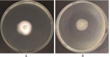
Fig.1
Growth forms of the isolatesA, Fungal colony morphology; B, Fungal colony backside"
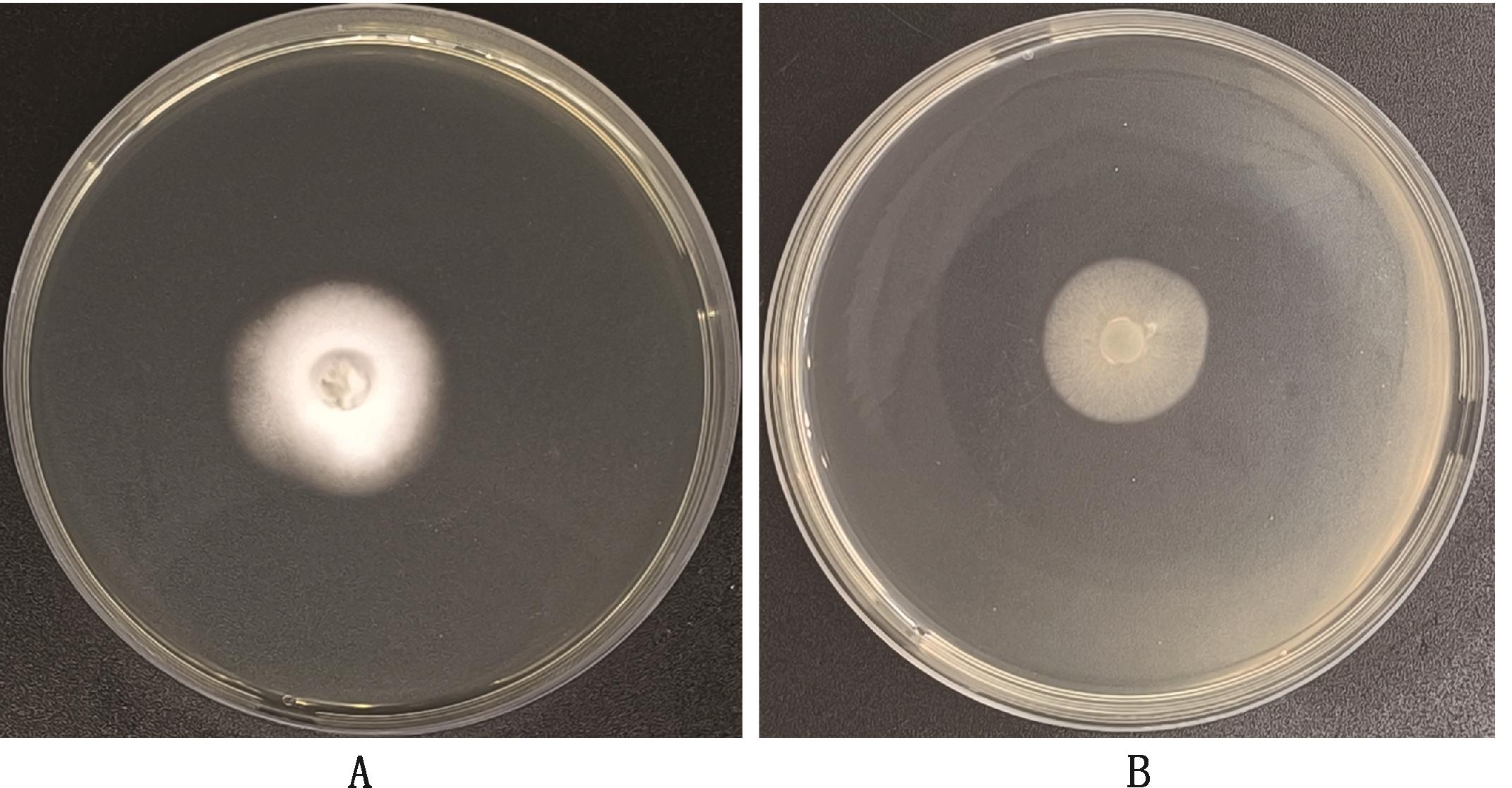
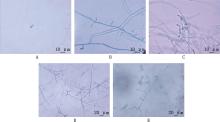
Fig.2
Microstructure of the isolatesA, Macroconidia (1 000×); B, Microconidia (1 000×); C, Thick-walled chaetospores(1 000×); D, Capsular spores (400×); E, Antler-like hyphae (400×)"
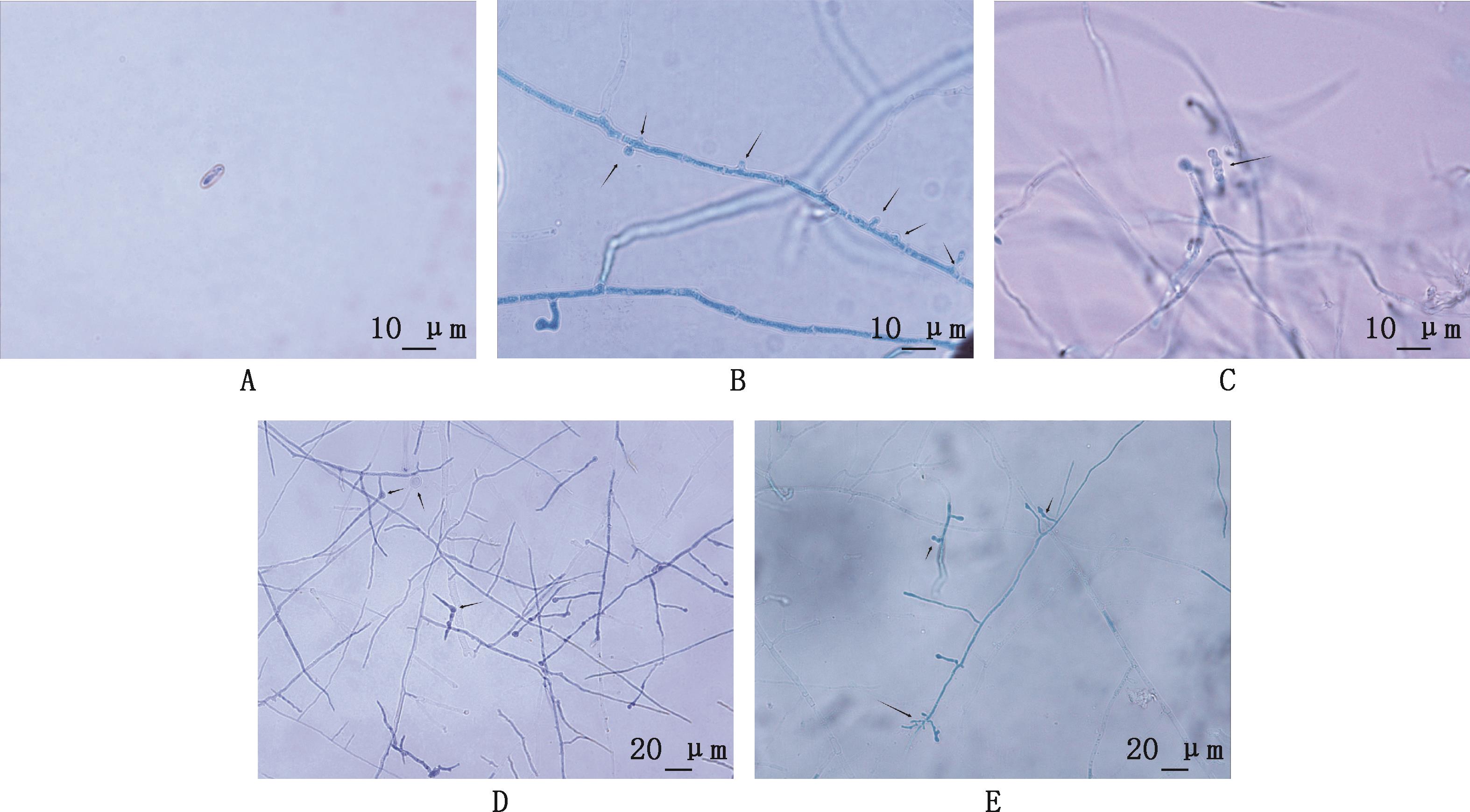
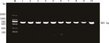
Fig.3
PCR amplification results of ITS rRNA gene sequence of the isolatesM, DL2000 DNA Marker; 1-10, The isolates"
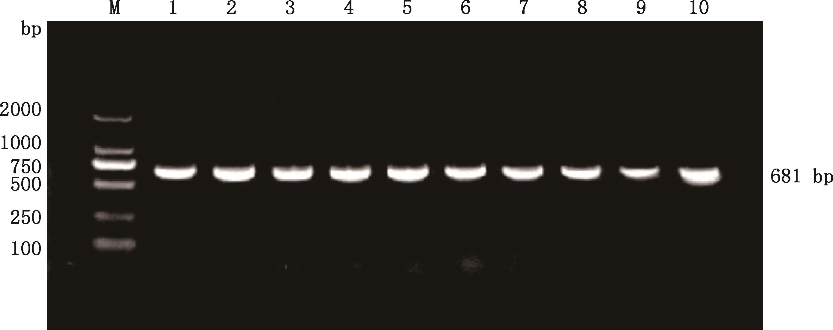

Fig.4
Phylogenetic tree based on ITS rRNA gene sequences"
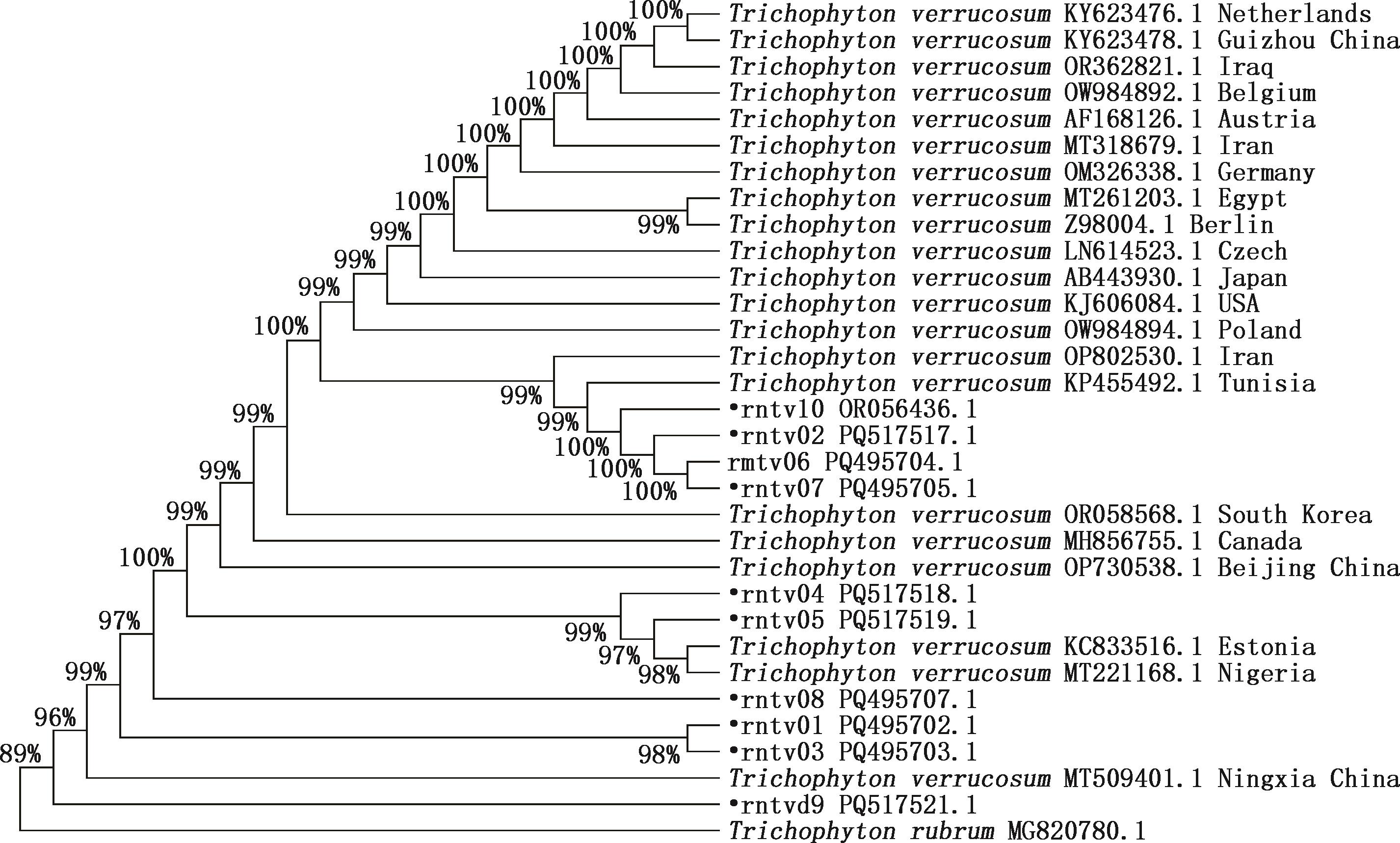
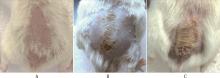
Fig.5
Clinical lesions in the skin of mice infected with T. verrucosumA, Normal uninoculated skin; B, 5 days after inculation; C, 14 days after inculation"
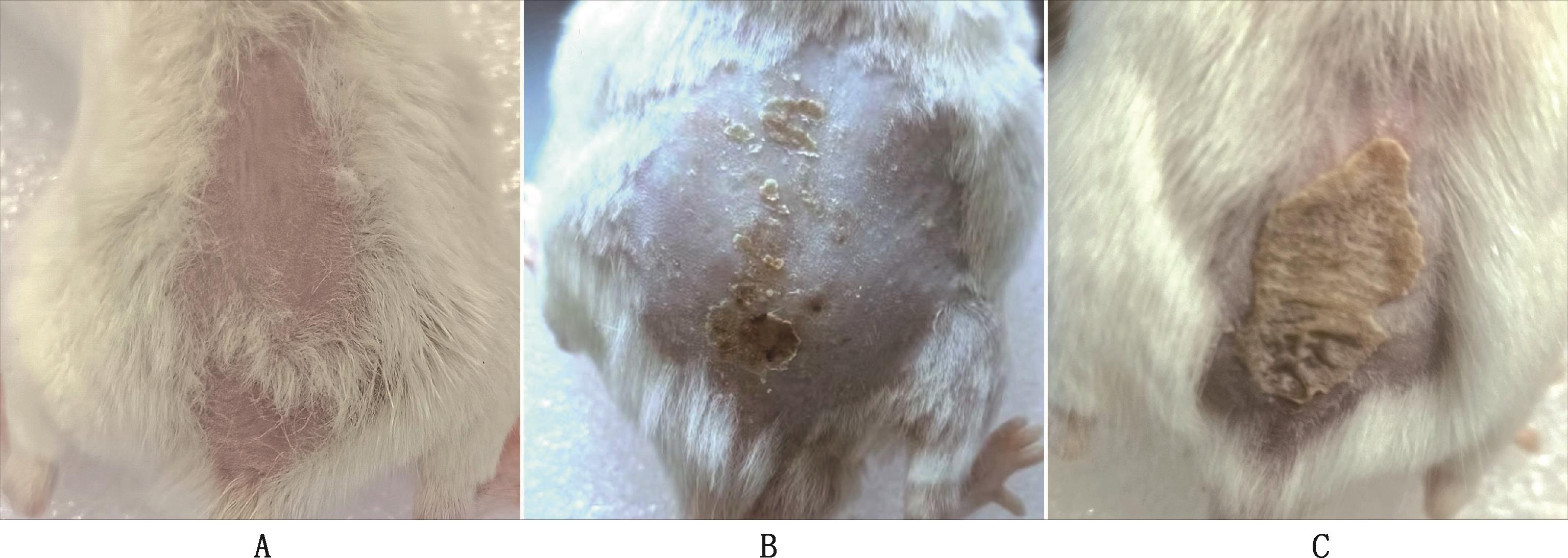
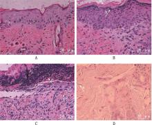
Fig.6
Histopathology observation of the skin of mice infected with T. verrucosum (400×)①A, Skin of mice in control group; B-D, Skin of mice in tested group. ② a, Dyskeratosis of epidermal stratum corneum; b, The granular layer and spinous layer of epidermis were thickened; c, Neutrophil and lymphocyte proliferation; d, Dermal and subcutaneous tissue congestion; e, Hyphal segments in the lesion"

Table 1
Drug susceptibility test results"
菌株 Strains | 最小抑菌浓度 MIC/(μg/mL) | ||||||
|---|---|---|---|---|---|---|---|
两性霉B AMB | 环吡酮胺 CPX | 伊曲康唑 ITR | 酮康唑 KTZ | 氟康唑 FCA | 5-氟胞嘧啶 5-FC | 特比萘芬 TEF | |
| rntv01 | 8(I) | 16(I) | 4(I) | 256(R) | 256(R) | >256(R) | >256(R) |
| rntv02 | 8(I) | 16(I) | 64(R) | 256(R) | 256(R) | >256(R) | >256(R) |
| rntv03 | 8(I) | 16(I) | 64(R) | 64(R) | 128(R) | >256(R) | >256(R) |
| rntv04 | 8(I) | 16(I) | 16(I) | 64(R) | 128(R) | >256(R) | >256(R) |
| rntv05 | 8(I) | 4(I) | 4(I) | 128(R) | 256(R) | >256(R) | >256(R) |
| rntv06 | 8(I) | 16(I) | 16(I) | 64(R) | 256(R) | >256(R) | >256(R) |
| rntv07 | 4(I) | 8(I) | 16(I) | 64(R) | 128(R) | >256(R) | >256(R) |
| rntv08 | 8(I) | 8(I) | 16(I) | 128(R) | 128(R) | >256(R) | >256(R) |
| rntv09 | 8(I) | 8(I) | 2(S) | 64(R) | 128(R) | >256(R) | >256(R) |
| rntv10 | 64(R) | 16(I) | 1(S) | 2(S) | 256(R) | 32(I) | 64(R) |
| [1] | 王建昌, 姜彦芬, 王 珅, 等. 奶牛疣状毛癣菌的分离鉴定及其耐药性分析[J]. 中国动物检疫, 2016, 33(1):26-29. |
| WANG J C, JIANG Y F, WANG S, et al. Isolation and identiifcation of Trichophyton verrucosum in cows and analysis on vitro drug resistance[J]. China Animal Health Inspection, 2016, 33(1): 26-29. (in Chinese) | |
| [2] | 崔鹏博. 奶牛皮肤真菌病病原分离及抗真菌药物的筛选[D].哈尔滨: 东北农业大学,2008. |
| CUI P B. Separate and verification of cows’dermatophytosis and screening of antifungal agent[D]. Harbin: Northeast Agricultural University, 2008. (in Chinese) | |
| [3] | 马 颖, 尤德渊. 某牧场人畜共患皮肤癣菌病病原菌调查[J]. 中国麻风皮肤病杂志, 2012, 28(7):467-469. |
| MA Y, YOU D Y. Investigation of the zoonotic dernatophytosis in a pasture[J]. China Journal of Leprosy and Skin Diseases, 2012, 28(7): 467-469. (in Chinese) | |
| [4] | 吴润标, 李前勇, 李 奥, 等. 山羊疣状毛癣菌鉴定与敏感天然药物的体外筛选试验[J]. 吉林农业大学学报, 2018, 40(3):358-363. |
| WU R B, LI Q Y, LI A, et al. Identification of Trichophyton verrucosum of goats and screening of sensitive natural medicine in vitro [J]. Journal of Jilin Agricultural University, 2018, 40(3): 358-363. (in Chinese) | |
| [5] | 尚盼盼, 周亚斌, 汪 旸, 等. 疣状毛癣菌致母女共患体癣分析[J]. 中国麻风皮肤病杂志, 2019, 35(4):225-228. |
| SHANG P P, ZHOU Y B, WANG Y, et al. Clinical and laboratory research on the tinea corporis on one mother and her daughter caused by Trichophyton verrucosum [J]. China Journal of Leprosy and Skin Diseases, 2019, 35(4): 225-228. (in Chinese) | |
| [6] | GUO Y, GE S, LUO H, et al. Occurrence of Trichophyton verrucosum in cattle in the Ningxia Hui autonomous region, China[J]. BMC Veterinary Research, 2020, 16(1):187. |
| [7] | KIM S J. Mycological features of Trichophyton verrucosum isolated in cattle[J]. Journal of Experimental & Biomedical Sciences, 2019, 25(4):367-371. |
| [8] | ILHAN Z. Isolation of dermatophytes from cattle, sheep, goats and Van cats in Van and its around[J]. Yüzüncü yıl Üniversitesi Veteriner Fakültesi Dergisi, 2015, 26(1):1-5. |
| [9] | AGNETTI F, RIGHI C, SCOCCIA E, et al. Trichophyton verrucosum infection in cattle farms of Umbria (Central Italy) and transmission to humans[J]. Mycoses, 2014, 57(7):400-405. |
| [10] | GNAT S, ŁAGOWSKI D, NOWAKIEWICZ A, et al. The host range of dermatophytes, it is at all possible? Phenotypic evaluation of the keratinolytic activity of Trichophyton verrucosum clinical isolates[J]. Mycoses, 2019, 62(3):274-283. |
| [11] | NÉJI S, MAKNI F, CHEIKROUHOU F, et al. Dermatomycosis due to Trichophyton verrucosum in Sfax-Tunisia[J]. Journal Demycologie Medicale, 2011, 21(3):198-201. |
| [12] | AHMED S, ISMAIL M, ALBIRAIR M, et al. Fungal infections in Sudan: An underestimated health problem[J]. PLoS Neglected Tropical Diseases, 2023, 17(9):e0011464. |
| [13] | DALIS J S, KAZEEM H M, KWAGA J K P, et al. Prevalence and distribution of dermatophytosis lesions on cattle in Plateau State, Nigeria[J]. Veterinary World, 2019, 12(9):1484-1490. |
| [14] | GNAT S, ŁAGOWSKI D, DYLĄG M, et al. Modulation of ERG gene expression in fluconazole-resistant human and animal isolates of Trichophyton verrucosum [J]. Brazilian Journal of Microbiology, 2021, 52(4):2439-2446. |
| [15] | ŁAGOWSKI D, GNAT S, NOWAKIEWICZ A, et al. Comparison of in vitro activities of 11 antifungal agents against Trichophyton verrucosum isolates associated with a variety hosts and geographical origin[J]. Mycoses, 2020, 63(3):294-301. |
| [16] | 宋秋荷, 周会祥, 赖雪花. 铜圈小培养鉴定真菌菌种的临床应用[J]. 九江学院学报(自然科学版), 2013, 28(1):76-77. |
| SONG Q H, ZHOU H X, LAI X H. Clinical application of copper-ring micro-culture in identifying fungal strains[J]. Journal of Jiujiang University (Natural Sciences), 2013, 28(1): 76-77. (in Chinese) | |
| [17] | 李俊树. 四川山鹧鸪肠道菌群的多样性及潜在病原体调查[D]. 成都: 四川农业大学,2023. |
| LI J S. Investigation on intestinal microflora diversityand potential pathogens of Arborophila rufipectus [D]. Chengdu: Sichuan Agricultural University, 2023. (in Chinese) | |
| [18] | 李 玲, 尤德渊. 疣状毛癣菌感染豚鼠模型的构建[J]. 中华皮肤科杂志, 2012, 45(8):582-583. |
| LI L, YOU D Y. Establishment of a guinea pig model of dermatophytosis caused by Trichophyton verrucosum [J]. Chinese Journal of Dermatology, 2012, 45(8): 582-583. (in Chinese) | |
| [19] | 刘 月. 兔须癣毛癣菌的鉴定及快速诊断方法的建立[D]. 邯郸: 河北工程大学,2016. |
| LIU Y. The identification and the establish of diagnostic method of Trichpohyton mentagrophyte in rabbits[D]. Handan: Hebei University of Engineering, 2016. (in Chinese) | |
| [20] | 刘 红. 磷脂酶A2(PLA2)基因对大熊猫源阿萨希毛孢子菌耐药性的影响[D]. 成都: 四川农业大学,2023. |
| LIU H. Influence of phospholipase A2 (PLA2) gene on the drug resistance of Trichosporon asahii isolated from giant pandas[D]. Chengdu: Sichuan Agricultural University, 2023. (in Chinese) | |
| [21] | SIOPI M, PACHOULIS I, LEVENTAKI S, et al. Evaluation of the Vitek 2 system for antifungal susceptibility testing of Candida auris using a representative international panel of clinical isolates: Overestimation of amphotericin B resistance and underestimation of fluconazole resistance[J]. Journal of Clinical Microbiology, 2024, 62(4): e01528-23. |
| [22] | 葛 松, 蒋 万, 何生虎, 等. 宁夏肉牛皮肤病原真菌的分离与鉴定[J]. 中国农业科学, 2015, 48(14):2876-2883. |
| GE S, JIANG W, HE S H, et al. Isolation and identification of dermatophytes from beef cattle in Ningxia[J]. Scientia Agricultura Sinica, 2015, 48(14): 2876-2883. (in Chinese) | |
| [23] | 王建昌, 姜彦芬, 王 珅, 等. 进口奶牛皮肤真菌病病原疣状毛癣菌的分离鉴定[J]. 西北农业学报, 2015, 24(7):1-5. |
| WANG J C, JIANG Y F, WANG S, et al. Isolation and identification of Trichophyton verrucosum from ringworm skin lesions of imported dairy cows[J]. Acta Agriculturae Boreali-occidentalis Sinica, 2015, 24(7): 1-5. (in Chinese) | |
| [24] | KHOSRAVI A, MANSOURI P, NIKAEIN D, et al. Severe tinea corporis due to Trichophyton verrucosum mimicking discoid lupus erythematosus[J]. Journal of Medical Mycology, 2012, 22(1):92-95. |
| [25] | O’GORMAN S M, BRITTON D J, COLLINS P M. An uncommon dermatophyte infection: Two cases of cutaneous infection with Trichophyton verrucosum [J]. Clinical and Experimental Dermatology, 2015, 40(4):395-398. |
| [26] | JIANG Y, ZHAN P, AL-HATMI A M S, et al. Extensive tinea capitis and corporis in a child caused by Trichophyton verrucosum [J]. Journal de Mycologie Medicale, 2019, 29(1):62-66. |
| [27] | GNAT S, ŁAGOWSKI D, NOWAKIEWICZ A, et al. Detection and identification of dermatophytes based on currently available methods-A comparative study[J]. Journal of Applied Microbiology, 2021, 130(1):278-291. |
| [28] | NENOFF P, ERHARD M, SIMON J C, et al. MALDI-TOF mass spectrometry—A rapid method for the identification of dermatophyte species[J]. Medical Mycology, 2012, 51(1):17-24. |
| [29] | UMITZHANOV M, ABDIRAMANOVA B, ABUTALIP A, et al. Comparative assessment of regulated methods and PCR in the diagnosis of trichophytosis in veterinary mycology[J]. Open Veterinary Journal, 2024, 13(12):1614. |
| [30] | VIJAYAKUMAR R, VIJAYARAMAN R S, HEMANTH V, et al. Molecular strain typing of Trichophyton mentagrophytes (T. mentagrophytes var. interdigitale) using non-transcribed spacer region as a molecular marker[J]. Directory of Open Access Journals, 2017, 146(5):636-641. |
| [31] | CIESIELSKA A, STĄCZEK P. A new molecular marker for species-specific identification of Microsporum canis [J]. Brazilian Journal of Microbiology, 2020, 51(4):1505-1508. |
| [32] | 吴润标,李小燕,李 奥,等. 牛羊皮肤癣菌病的研究进展[J]. 中国兽医杂志, 2016,52(11):79-81. |
| WU R B, LI X X, LI A, et al. Research progress of dermatophytosis in cattle and sheep[J]. Chinese Journal of Veterinary Medicine, 2016, 52(11):79-81. (in Chinese) | |
| [33] | SONG X, YUAN W, LAI KM, et al. In vivo antifungal activity of dipyrithione against Trichophyton rubrum on guinea pig dermatophytosis models[J]. Biomedicine & Pharmacotherapy, 2018, 108:558-564. |
| [34] | MIKAEILI A, MODARESI M, KARIMI I, et al. Antifungal activities of Astragalus verus Olivier. against Trichophyton verrucosum on in vitro and in vivo guinea pig model of dermatophytosis[J]. Mycoses, 2011, 55(4):318-325. |
| [35] | LEE H G, BOK E Y, JUNG Y H, et al. Antifungal activity of aminopyrrolnitrin against Trichophyton verrucosum in a guinea pig model of dermatophytosis[J]. Mycoses, 2024, 67(5):e13748. |
| [36] | 李丽丽, 崔 英, 王丽梅, 等. 疣状毛癣菌的体外抗真菌药物敏感性测试的研究[J]. 中国医师杂志, 2015, 17(11):1739-1740. |
| LI L L, CUI Y, WANG L M, et al. Study on in vitro antifungal susceptibility testing of Trichophyton verrucosum [J]. Journal of Chinese Physicians, 2015, 17(11): 1739-1740. (in Chinese) | |
| [37] | 杨朝位. 牛皮肤真菌病的科学防控分析[J]. 吉林畜牧兽医, 2024, 45(12):115-117. |
| YANG C W. Analysis of scientific prevention and control of bovine dermatomycosis[J]. Jilin Animal Husbandry and Veterinary Medicine, 2024, 45(12): 115-117. (in Chinese) |
| [1] | ZHANG Sui, CHENG Chunjie, YANG Chun, CHEN Mengjiao, CHENG Jing, ZHOU Linyi, LI Yongqing, WANG Xiaoying, LIU Wenxiao. Isolation, Identification and Pathogenicity Analysis of Dairy Cow-derived Klebsiella pneumoniae and Mannheimia haemolytica [J]. China Animal Husbandry & Veterinary Medicine, 2026, 53(2): 903-915. |
| [2] | ZHANG Min, LI Yingxiao, ZHANG Lulu, HE Shuhai, QU Zhehui, QIN Dongsheng, LIU Jicheng, JIAO Fengchao. Whole Genome Amplification and Phylogenetic Analysis of Duck Astrovirus Type 1 Xinyang Strain [J]. China Animal Husbandry & Veterinary Medicine, 2026, 53(2): 959-972. |
| [3] | ZHANG Xinxin, ZHANG Yue, CUI An, LI Jinru, ZHANG Yunjing, HE Lixia, YANG Guijun, REN Yudong, LI Guangxing. Isolation, Identification and Genetic Evolution Analysis of PEDV Epidemic Strains in Heilongjiang Province [J]. China Animal Husbandry & Veterinary Medicine, 2026, 53(1): 378-389. |
| [4] | LIAN Kaiqi, WANG Yuhang, MENG Xiangfeng, ZHANG Mingliang, ZHOU Lingling. Isolation, Identification and Drug Resistance Analysis of Pathogenic Escherichia coli from Pavo cristatus [J]. China Animal Husbandry & Veterinary Medicine, 2026, 53(1): 448-458. |
| [5] | WANG Qilin, CAO Runlai, LIU Peiwen, LIU Ziyu, XUE Zhuqing, DU Haixia, ZHANG Yue, ZHANG Minjing, LI Yacong, WANG Weiyang, CONG Yanfang, WANG Xiaoxu, LIU Zhijie. Isolation, Identification and Biological Characteristics Analysis of Bordetella bronchiseptica from Raccoon dog(Nyctereutes procyonoides) [J]. China Animal Husbandry & Veterinary Medicine, 2026, 53(1): 489-498. |
| [6] | LI Fuxiang, SONG Jianling, LI Zhanhong. Isolation, Identification and Biological Characterization of Actinobacillus pleuropneumoniae Serotype 7 from Pigs in Yunnan [J]. China Animal Husbandry & Veterinary Medicine, 2026, 53(1): 499-508. |
| [7] | ZHAO Xuejian, CHEN Xiaoliang, FENG Xiaowei, ZHANG Kang, MA Xiaojun, LI Jianxi. Investigation of the Pathogen of Calf Diarrhea in Gansu Region and Isolation, Identification and Drug Resistance Analysis of Clostridium perfringens [J]. China Animal Husbandry & Veterinary Medicine, 2025, 52(8): 3927-3938. |
| [8] | ZHONG Guangzhi, LI Mengyue, TUO Shiyou, SHAN Gang, WANG Zixuan, LI Jing. Research Progress on the Causes of Embryonic Death and Abortion in Beef Cattle [J]. China Animal Husbandry & Veterinary Medicine, 2025, 52(8): 3999-4007. |
| [9] | DAI Lin, HUAI Zhifang, WANG Baowei, ZHANG Ming'ai, FAN Wenlei, ZHANG Jing, WANG Binghan, QI Guofeng. Isolation, Identification and Biological Properties and Safety of A Lactiplantibacillus plantarum Strain [J]. China Animal Husbandry & Veterinary Medicine, 2025, 52(7): 3070-3083. |
| [10] | LU Chong, WANG Yuyan, FU Han, LI Tongyang, MIAO Ronghao, LU Yabin, LI Jianlong, LIU Jianhua, GUO Qingyong, MAI Zhanhai, KUANG Ling. Isolation and Identification of the Main Pathogenic Bacteria of Aborted Yili Foals and Their Pathogenicity [J]. China Animal Husbandry & Veterinary Medicine, 2025, 52(7): 3344-3358. |
| [11] | ZHAO Yilong, TANG Na, JING Changhua, XU Qingqing, YIN Xiusheng, WANG Haiming, SUN Jing, LIN Panpan, DONG Lin, LIU Jishan, CAO Rongfeng. Isolation,Identification and Biological Characteristics of a Strain of Lumpy Skin Disease Virus [J]. China Animal Husbandry & Veterinary Medicine, 2025, 52(6): 2790-2799. |
| [12] | YANG Qinhong, YANG Jia, DUAN Wenjie, ZHANG Yongxian, ZHU Youshuai, YANG Shaohong, HE Cheng, YIN Hongbin, LI Suhua. Isolation and Identification of a High-temperature Tolerant Bacillus thermoamylovorans and Optimization of Its Enzyme Production Conditions [J]. China Animal Husbandry & Veterinary Medicine, 2025, 52(6): 2839-2850. |
| [13] | WU Peng, YUAN Yang, YANG Yunyun, FENG Yi, WANG Yan, YANG Ying, CHEN Jiangfeng, JIANG Haibo, WEN Ming. Diagnosis and Pathogen Identification of a Case of Mixed Infection of Duck Plague Virus and Pasteurella multocida in Breeding Goose [J]. China Animal Husbandry & Veterinary Medicine, 2025, 52(5): 2253-2265. |
| [14] | WU Yue, SHI Xingya, ZHANG Shuai, ZHAO Yunhuan, GUO Liming, ZUO Yuzhu, FAN Jinghui. Isolation and Identification of Porcine Pseudorabies Virus and Genetic Evolution Analysis of gE and gC Genes [J]. China Animal Husbandry & Veterinary Medicine, 2025, 52(5): 2266-2277. |
| [15] | YU Kun, ZHAO Jie, MA Qin, SHI Yanhong, ZHANG Xiao, LIU Zihan, ZHANG Xinting, WANG Jianhua, LI Yufeng. Isolation,Identification,Drug Resistance and Pathogenicity Analysis of Salmonella Enteritidis from Commercial Meat Ducks [J]. China Animal Husbandry & Veterinary Medicine, 2025, 52(5): 2353-2363. |
| Viewed | ||||||
|
Full text |
|
|||||
|
Abstract |
|
|||||