
China Animal Husbandry & Veterinary Medicine ›› 2026, Vol. 53 ›› Issue (1): 427-438.doi: 10.16431/j.cnki.1671-7236.2026.01.038
• Basic Veterinary Medicine • Previous Articles Next Articles
QI Sichao1,2( ), YU Haolin1, ZHANG Lingzhi1, HUA Yuping1, CHEN Leixiao1, WANG Huanan1, LI Jian1()
Received:2025-04-02
Online:2026-01-05
Published:2025-12-26
Contact:
LI Jian
E-mail:scqi@zju.edu.cn;lijiannp@zju.edu.cn
CLC Number:
QI Sichao, YU Haolin, ZHANG Lingzhi, HUA Yuping, CHEN Leixiao, WANG Huanan, LI Jian. Potential Mechanisms of Intestinal Mucosal Defense Against Secondary Injury in Laying Hens[J]. China Animal Husbandry & Veterinary Medicine, 2026, 53(1): 427-438.
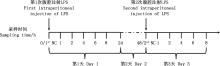
Fig.1
Group setting and treatments"
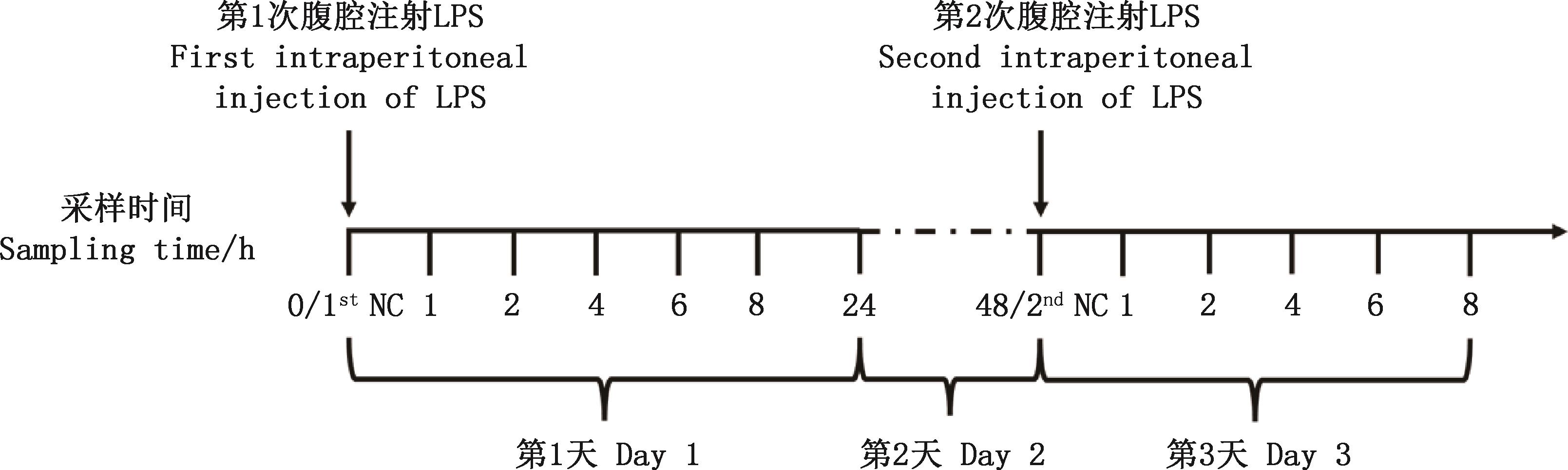
Table 1
Real-time quantitative PCR primer information"
基因 Genes | 引物序列 Primer sequences (5'→3') | 产物长度 Product length/bp | GenBank登录号 GenBank accession No. |
|---|---|---|---|
| GAPDH | F: GATGGGTGTCAACCATGAGAAA | 116 | NM_204305.1 |
| R: CAATGCCAAAGTTGTCATGGA | |||
| Claudin-1 | F: GTGTTTGTTGCTGTGACGGG | 153 | NM_001013611.2 |
| R: AGCCACTCTGTTGCCATACC | |||
| Ocln | F: ACAGCCCTCAATACCAGGATGTG | 133 | XM_052699761.1 |
| R: ACCATGCGCTTGATGTGGAA | |||
| Muc2 | F: TACTTCACCTTCAACCATTACAAC | 161 | NM_001318434.1 |
| R: CATAGTCACCACCATCTTCTTCA | |||
| C5 | F: CCTCCATCCAGGCCTTTCAG | 85 | XM_025141495.3 |
| R: GCCATCTGAAGGGAAAGAACC | |||
| TLR4 | F: GGATCTTTCAAGGTGCCACA | 134 | NM_001030693.2 |
| R: GCGACGTTAAGCCATGGAAG | |||
| IL-4 | F: TGCTTACAGCTCTCAGTGCC | 79 | NM_001007079.2 |
| R: TCTTGACGCAGGAAACCTCTC |
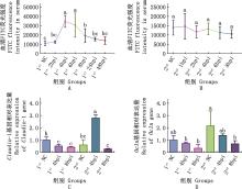
Fig.2
Effect of two times LPS induced injuries on mechanical barrier of intestinal mucosa①A and B, The changes in serum fluorescence intensity after the first and second LPS injections following the administration of FITC glucan, respectively; C and D, Relative expression of tight junction protein Claudin-1 and Ocln genes in duodena, respectively. ②Values with different letter superscripts mean significant difference (P<0.05);While with the same letter superscripts mean no significant difference (P>0.05). The same as below"

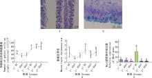
Fig.3
Effect of two times LPS induced injuries on chemical barrier of intestinal mucosaA and C, Schematic diagrams of goblet cell staining and corresponding quantitative results, respectively; B and D, Schematic diagrams of mucus layer staining and corresponding quantitative results, respectively; E, Relative expression level of Muc2 gene in duodena"
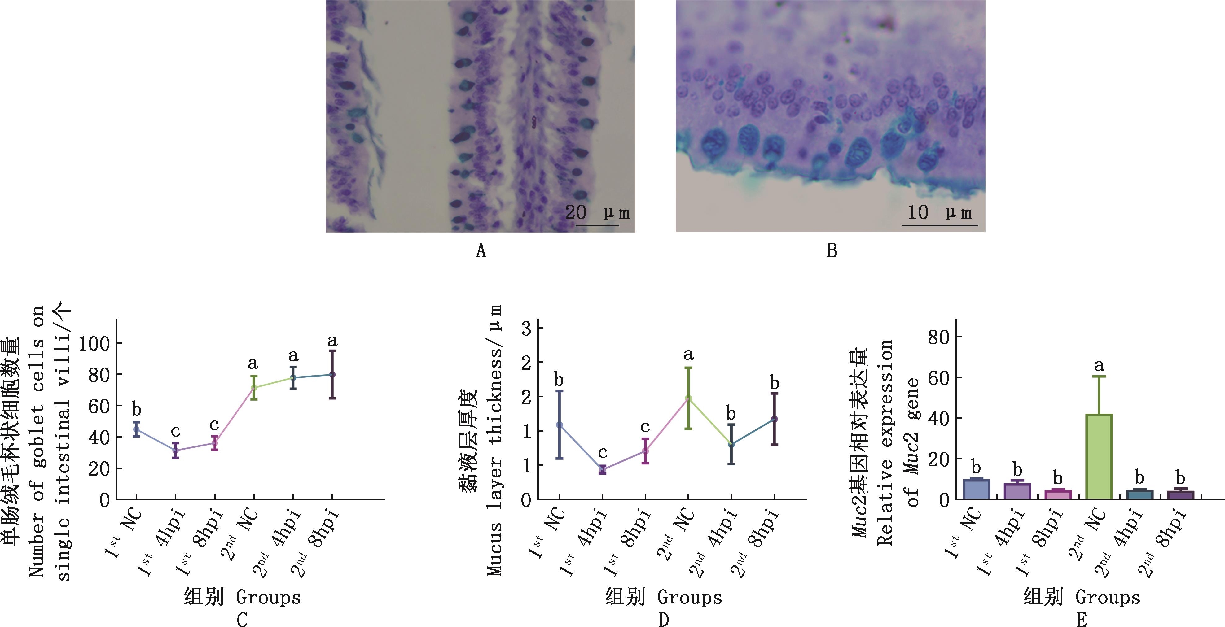
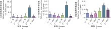
Fig.4
Effect of two times LPS induced injuries on immune barrier of intestinal mucosa"

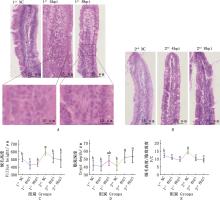
Fig.5
Effect of two times LPS induced injuries on morphology of intestinal mucosaA and B, H&E staining results of intestinal villi during the 1st and the 2nd injury; C and D, Duodenal villus height and crypt depth detection result, respectively; E, Statistical results of duodenal villus height/crypt depth"
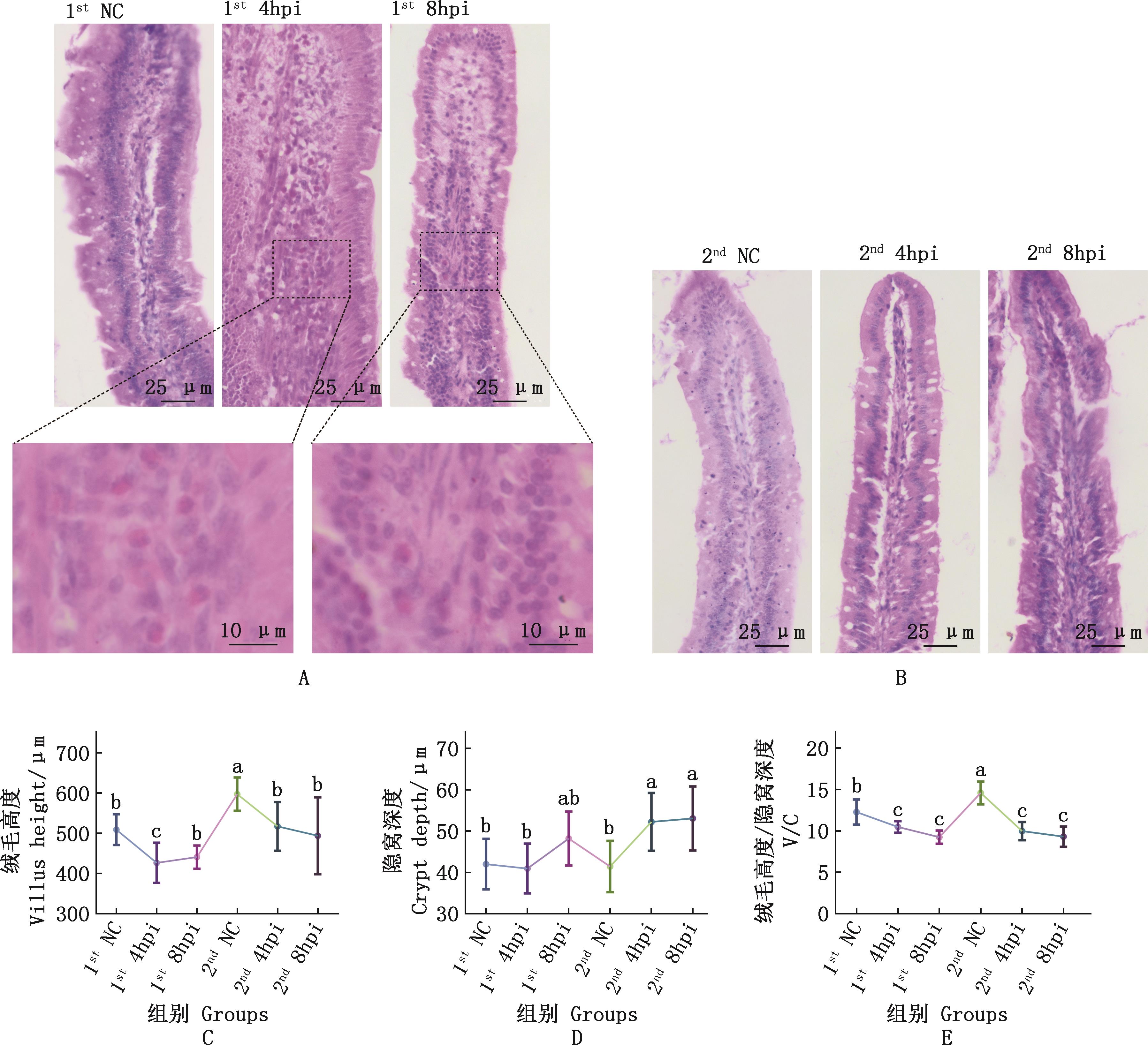
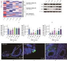
Fig.6
Effect of two times LPS induced injuries on the activity of ISCsA, Heatmap of genes related to intestinal crypt ISCs markers and ISCs regulatory factors after two times LPS injections; B-E, Western blotting and the statistical analysis results of LGR5, HOPX and PCNA proteins expression in duodenal crypt; F, Immunofluorescence staining of LGR5 protein in duodenum; G, The enlarged image of the boxed area in fig.F; H, Negative control of duodenal immunofluorescence staining"

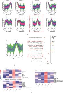
Fig.7
Transcriptome time-seres analysis of intestinal cryptsA, Transcriptome time-seres analysis results; B, The magnified image of Cluster 7; C, Enrichment of the top 10 KEGG pathways of genes in Cluster 7; D, Expression heatmap of genes in Cluster 7 which enriched in KEGG pathway mucin type O-glycan biosynthesis, steroid hormone biosynthesis and intestinal immune network for IgA production"


Fig.8
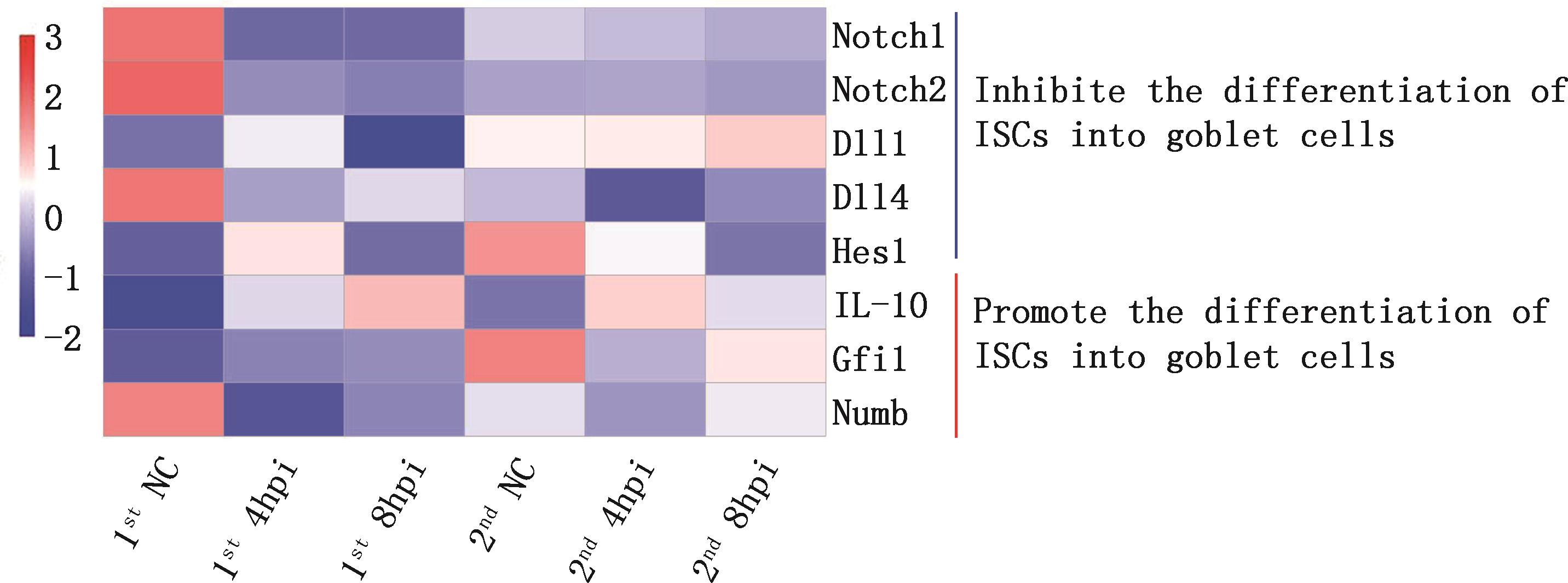
Heatmap of genes regulating the differentiation of ISCs into goblet cells"
| [1] | NAIK S, LARSEN S B, GOMEZ N C, et al. Inflammatory memory sensitizes skin epithelial stem cells to tissue damage[J]. Nature, 2017,550(7677):475-480. |
| [2] | LYNCH T J, ANDERSON P J, ROTTI P G, et al. Submucosal gland myoepithelial cells are reserve stem cells that can regenerate mouse tracheal epithelium[J]. Cell Stem Cell, 2018,22(5):653-667. |
| [3] | YU L, XIE X, JIANG K, et al. Paneth cells mediated the response of intestinal stem cells at the early stage of intestinal inflammation in the chicken[J]. Poultry Science, 2021,100(2):615-622. |
| [4] | BU P, WANG L, CHEN K Y, et al. A miR-34a-numb feedforward loop triggered by inflammation regulates asymmetric stem cell division in intestine and colon cancer[J]. Cell Stem Cell, 2016,18(2):189-202. |
| [5] | MA Y, LANG X, YANG Q, et al. Paeoniflorin promotes intestinal stem cell-mediated epithelial regeneration and repair via PI3K-Akt-mTOR signalling in ulcerative colitis[J]. International Immunopharmacology, 2023,119:110247. |
| [6] | BHANJA P, NORRIS A, GUPTA-SARAF P, et al. BCN057 induces intestinal stem cell repair and mitigates radiation-induced intestinal injury[J]. Stem Cell Research & Therapy, 2018,9(1):26. |
| [7] | TIAN H, BIEHS B, WARMING S, et al. A reserve stem cell population in small intestine renders Lgr5-positive cells dispensable[J]. Nature, 2011,478(7368):255-259. |
| [8] | STEWART A S, SCHAAF C R, LUFF J A, et al. HOPX+ injury-resistant intestinal stem cells drive epithelial recovery after severe intestinal ischemia[J]. American Journal of Physiology-Gastrointestinal and Liver Physiology, 2021,321(5):G588-G602. |
| [9] | LI N, ZHANG Y, NEPAL N, et al. Dental pulp stem cells overexpressing hepatocyte growth factor facilitate the repair of DSS-induced ulcerative colitis[J]. Stem Cell Research & Therapy, 2021,12(1):30. |
| [10] | SAILAJA B S, HE X C, LI L. The regulatory niche of intestinal stem cells[J]. Journal of Physiology-London, 2016,594(17):4827-4836. |
| [11] | YANG S, YU M. Role of goblet cells in intestinal barrier and mucosal immunity[J]. Journal of Inflammation Research, 2021,14:3171-3183. |
| [12] | VENUGOPAL S, ANWER S, SZASZI K. Claudin-2: Roles beyond permeability functions[J]. International Journal of Molecular Sciences, 2019,20(22):5655. |
| [13] | VUONG C N, MULLENIX G J, KIDD M T, et al. Research note: Modified serum fluorescein isothiocyanate dextran (FITC-d) assay procedure to determine intestinal permeability in poultry fed diets high in natural or synthetic pigments[J]. Poultry Science, 2021,100(6):101138. |
| [14] | KANG J, ZHOU Y, ZHU C, et al. Ginsenoside Rg1 mitigates porcine intestinal tight junction disruptions induced by LPS through the p38 MAPK/ NLRP3 inflammasome pathway[J]. Toxics, 2022,10(6):285. |
| [15] | GUO C, GUO D, FANG L, et al. Ganoderma lucidum polysaccharide modulates gut microbiota and immune cell function to inhibit inflammation and tumorigenesis in colon[J]. Carbohydrate Polymers, 2021,267:118231. |
| [16] | MA J, ZHANG J, WANG Y, et al. Modified Gegen Qinlian decoction ameliorates DSS-induced chronic colitis in mice by restoring the intestinal mucus barrier and inhibiting the activation of gammadeltaT17 cells[J]. Phytomedicine, 2023,111:154660. |
| [17] | BERGSTROM K S, KISSOON-SINGH V, GIBSON D L, et al. Muc2 protects against lethal infectious colitis by disassociating pathogenic and commensal bacteria from the colonic mucosa[J]. PLoS Pathogens, 2010,6(5):e1000902. |
| [18] | LAUMONNIER Y, KORKMAZ R U, NOWACKA A A, et al. Complement-mediated immune mechanisms in allergy[J]. European Journal of Immunology, 2023,53(10):e2249979. |
| [19] | GUO J, ZHANG Q Y, XU L, et al. Icariin ameliorates LPS-induced acute lung injury in mice via complement C5a-C5aR1 and TLR4 signaling pathways[J]. International Immunopharmacology, 2024,131:111802. |
| [20] | RADPOUR M, KHOSHKROODIAN B, ASGARI T, et al. Interleukin 4 reduces brain hyperexcitability after traumatic injury by downregulating TNF-alpha, upregulating IL-10/TGF-beta, and potential directing macrophage/microglia to the M2 anti-inflammatory phenotype[J]. Inflammation, 2023,46(5):1810-1831. |
| [21] | GAO S, ZHOU J, LIU N, et al. Curcumin induces M2 macrophage polarization by secretion IL-4 and/or IL-13[J]. Journal of Molecular and Cellular Cardiology, 2015,85:131-139. |
| [22] | YU L, QI S, WEI G, et al. Kruppel-like factor 5 activates chick intestinal stem cell and promotes mucosal repair after impairment[J]. Cell Cycle, 2023,22(19):2142-2160. |
| [23] | MORALES R A, RABAHI S, DIAZ O E, et al. Interleukin-10 regulates goblet cell numbers through Notch signaling in the developing zebrafish intestine[J]. Mucosal Immunology, 2022,15(5):940-951. |
| [24] | MEURETTE O, MEHLEN P. Notch signaling in the tumor microenvironment[J]. Cancer Cell, 2018,34(4):536-548. |
| [25] | ZHANG S, ZHANG S, HOU Y, et al. Porcine Deltacoronavirus infection disrupts the intestinal mucosal barrier and inhibits intestinal stem cell differentiation to goblet cells via the Notch signaling pathway[J]. Journal of Virology, 2023,97(6):e0068923. |
| [26] | LI C, MA D, ZHOU H, et al. Effects of different doses lipopolysaccharides on the mucosal barrier in mouse intestine[J]. Research in Veterinary Science, 2020,133:75-84. |
| [27] | ALVARADO D M, CHEN B, ITICOVICI M, et al. Epithelial indoleamine 2,3-dioxygenase 1 modulates aryl hydrocarbon receptor and Notch signaling to increase differentiation of secretory cells and alter mucus-associated microbiota[J]. Gastroenterology, 2019,157(4):1093-1108. |
| [28] | AHMED S, MALDERA J A, KRUNIC D, et al. Fitness trade-offs incurred by ovary-to-gut steroid signalling in Drosophila [J]. Nature, 2020,584(7821):415-419. |
| [29] | VELARDI E, TSAI J J, HOLLAND A M, et al. Sex steroid blockade enhances thymopoiesis by modulating Notch signaling[J]. Journal of Experimental Medicine, 2014,211(12):2341-2349. |
| [1] | WANG Huixin, GAO Qingtao, LI Jiaheng, ZHANG Shunfen, ZHONG Ruqing, WANG Yang, CHEN Liang, ZHANG Hongfu. Effects of Lactobacillus plantarum Selenium on Tissue Morphology, Antioxidant Function and Inflammatory Indices in Ovary and Oviduct of Laying Hens [J]. China Animal Husbandry & Veterinary Medicine, 2026, 53(2): 682-691. |
| [2] | HAO Zhilei, ZHANG Hongrui, MA Yun. Effects of Different Oil Sources on Laying Performance, Egg Quality, and Fatty Acid Composition of Egg Yolk [J]. China Animal Husbandry & Veterinary Medicine, 2026, 53(2): 703-710. |
| [3] | ZOU Yinghao, WANG Yong, SUN Lihua, CHEN Xing, WANG Jie, LIU Guohua, WANG Kun, ZHENG Aijuan, ZHENG Shugui. The Effects of Different Levels of Fat Powder on the Egg-laying Performance and Egg Quality of Hy-Line Brown Laying Hens [J]. China Animal Husbandry & Veterinary Medicine, 2026, 53(1): 233-244. |
| [4] | WANG Yuyan, LI Menglin, CAO Qingyun, ZHOU Qiaoyi, ZHANG Xingyue, XIAO Yaqi, HUANG Shirui, ZHANG Zhiying, HU Tiesheng, SHI Dayou. Effect of Femented Tea on Laying Performance of Laying Hens After Forced Moulting [J]. China Animal Husbandry & Veterinary Medicine, 2025, 52(8): 3574-3583. |
| [5] | BA Hongli, MA Ning. Research Progress on the Mechanism and Application of Bacillus subtilis in Regulating Intestinal Health of Laying Hens [J]. China Animal Husbandry & Veterinary Medicine, 2025, 52(8): 3976-3986. |
| [6] | LI Jianing, JIANG Zhihui, XING Yueteng, MA Shengming. Effects of 1, 8-cineole Supplementation in Diet on the Antioxidant Capacity of Laying Hens and Eggs [J]. China Animal Husbandry & Veterinary Medicine, 2025, 52(7): 3016-3030. |
| [7] | WANG Beibei, HAO Erying, ZHANG Haihua, ZHANG Haijun, QIU Kai, WU Shugeng. Effects of Dietary Organic Manganese-Iron-Copper-Zinc Compound Preparations on the Production Performance,Blood Biochemical Indices, Follicle Development and Tibia Quality of Laying Hens [J]. China Animal Husbandry & Veterinary Medicine, 2025, 52(5): 2012-2022. |
| [8] | LAN Zhongqi, LI Peng, SHEN Shuang, JIANG Jinfeng, QIN Hongdeng, XIANG Yuting, WANG Jiaxiang. Effects of Dietary Epidermal Growth Factor Supplementation on Morphology, Hormone Content and Expression of Apoptosis Factors of Ovaries in Laying Hens [J]. China Animal Husbandry & Veterinary Medicine, 2025, 52(5): 2035-2044. |
| [9] | WANG Yuanzhuo, LIU Tong, WANG Guizhen, HAN Kunliang, GUO Hua. Effects of Compound Chinese Herb Ultrafine Powder on Immune Function and Related Gene Expression of Xinyang Black-feathered Laying Hens [J]. China Animal Husbandry & Veterinary Medicine, 2025, 52(12): 5661-5669. |
| [10] | ZHANG Ming, GU Yan, AO Xiang, ZHOU Jianchuan. Research Progress on Application of Phytase in Low-phosphorus Diets of Laying Hens [J]. China Animal Husbandry & Veterinary Medicine, 2025, 52(10): 4627-4640. |
| [11] | QIU Kai, CHANG Xinyu, GAO Shan, ZHANG Haihua, ZHANG Haijun, WU Shugeng. Effects of Dietary Calcium Formate Supplementation on Production Performance and Egg Quality of Elderly Laying Hens [J]. China Animal Husbandry & Veterinary Medicine, 2024, 51(8): 3320-3328. |
| [12] | CHAN Yanzi, WANG Xinyue, LI Sihan, WANG Junkai, WANG Yuyan, HU Tiesheng, MO Guifen, LIANG Dehong, SHI Dayou. Effects of Compound Traditional Chinese Medicine on Production Performance and Reproductive Function of Forced Molting Laying Hens [J]. China Animal Husbandry & Veterinary Medicine, 2024, 51(3): 1151-1159. |
| [13] | HUANG Jing, ZHAO Na, GUO Wanzheng, JIN Feng, CHEN Fang, ZHU Wei, FAN Qiwen, DU Encun, TAO Wenjing, HUANG Shaowen, WEI Jintao. Effects of Feed Mulberry on Production Performance,Egg Quality and Intestinal Tissue Morphology of Laying Hens [J]. China Animal Husbandry & Veterinary Medicine, 2024, 51(2): 540-548. |
| [14] | GUO Fangchao, JIA Ling, CHEN Wenfeng, CHEN Liang, MU Qingqing, XU Shuying, WANG Yongjuan. Effects of Fermented Soybean Meal on the Production Performance,Antioxidant Capacity and Gut Microbiota Diversity of Laying Hens in the Late Laying Period [J]. China Animal Husbandry & Veterinary Medicine, 2024, 51(12): 5244-5253. |
| [15] | QIU Kai, GUAN Xiaofeng, SANG Yang, LIU Zhiyun, LIU Guohua. Effects of Lactobacillus acidophilus Post-Biotic on Production Performance, Blood Indexes and Egg Quality of Laying Hens in the Initial Laying Stage [J]. China Animal Husbandry & Veterinary Medicine, 2024, 51(11): 4824-4832. |
| Viewed | ||||||
|
Full text |
|
|||||
|
Abstract |
|
|||||