
China Animal Husbandry & Veterinary Medicine ›› 2026, Vol. 53 ›› Issue (1): 107-118.doi: 10.16431/j.cnki.1671-7236.2026.01.010
• Review • Previous Articles Next Articles
RUAN Qianhua1( ), LIN Jiayan2, LIN Shiqi1, ZHAO Jin1, LIU Chuandun2, LI Ying1, ZHANG Hui1, ZHANG Yuan1()
Revised:2025-05-27
Online:2026-01-05
Published:2025-12-26
Contact:
ZHANG Yuan
E-mail:1093741359@qq.com;zhangyuan@scau.edu.cn
CLC Number:
RUAN Qianhua, LIN Jiayan, LIN Shiqi, ZHAO Jin, LIU Chuandun, LI Ying, ZHANG Hui, ZHANG Yuan. Advances in Pathological Mechanisms and Treatment of Canine Myxomatous Mitral Valve Disease[J]. China Animal Husbandry & Veterinary Medicine, 2026, 53(1): 107-118.

Fig.1
Structurally normal mitral valve (A) and structurally abnormal (MMVD) mitral valve (B)"

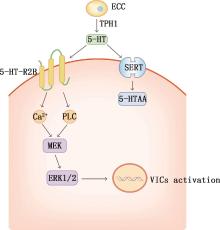
Fig.2
Mechanism of activation of VICs by 5-HT"

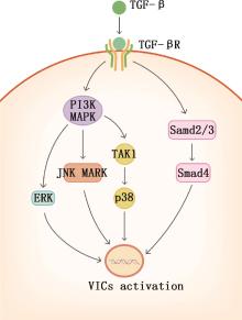
Fig.3
Mechanism of activation of VICs by TGF-β"
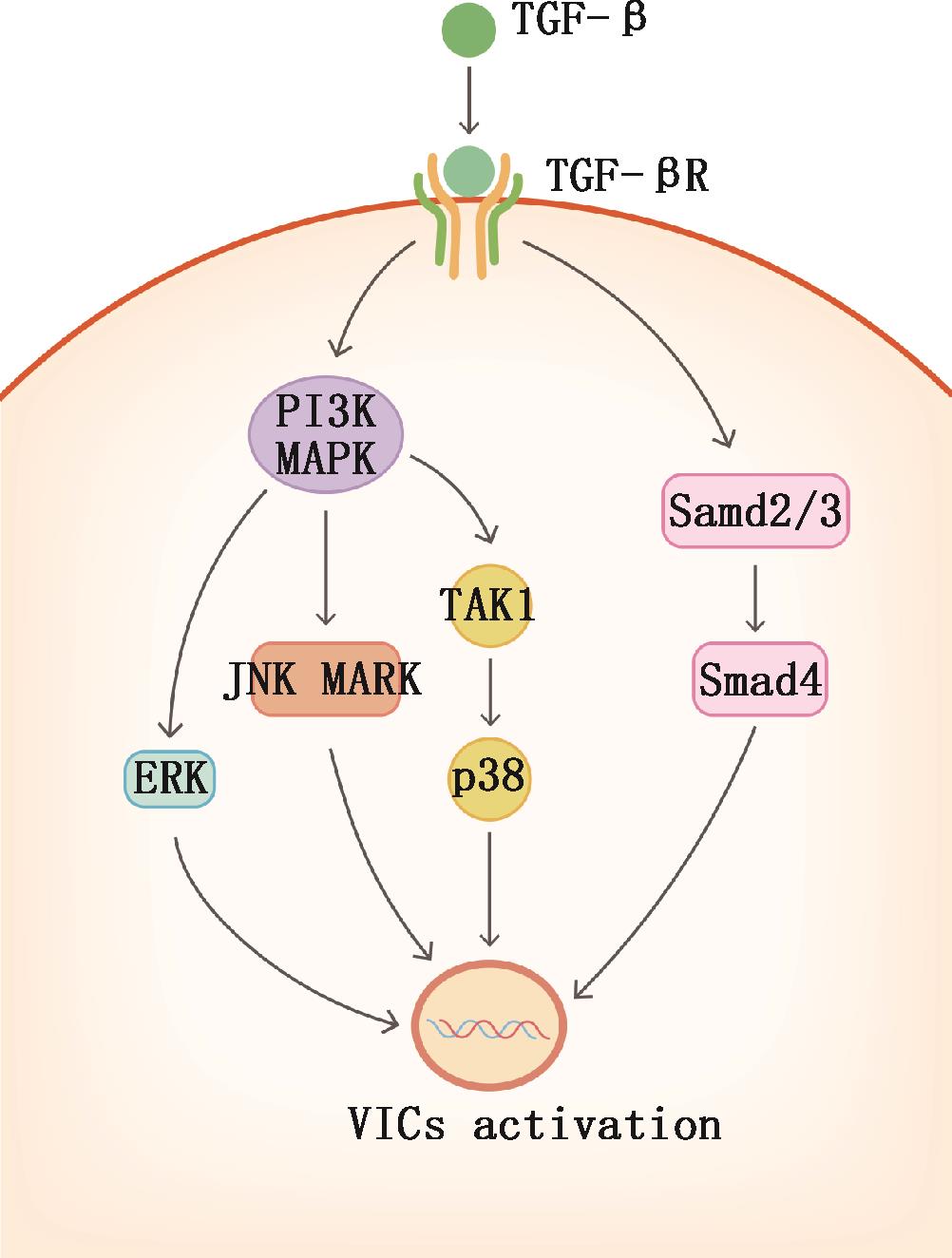
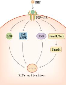
Fig.4
Mechanism of activation of VICs by BMP"

| [1] | KEENE B W, ATKINS C E, BONAGURA J D, et al. ACVIM consensus guidelines for the diagnosis and treatment of myxomatous mitral valve disease in dogs[J]. Journal of Veterinary Internal Medicine, 2019,33(3):1127-1140. |
| [2] | ADLER E, TIDHOLM A. Prevalence of mitral valve regurgitation in 102 asymptomatic Chinese Crested dogs[J]. Journal of Veterinary Cardiology, 2023,46:55-61. |
| [3] | 陈淑敏,曾丽薇,陈宇,等.犬心脏二尖瓣退行性疾病研究进展[J]. 中国畜牧兽医, 2024,51(7):3215-3223. |
| CHEN S M, ZENG L W, CHEN Y, et al. Research progress on mitral degenerative disease of dogs[J]. China Animal Husbandry & Veterinary Medicine, 2024,51(7):3215-3223. (in Chinese) | |
| [4] | FISHBEIN G A, FISHBEIN M C. Mitral valve pathology[J]. Current Cardiology Reports, 2019,21(7):61. |
| [5] | MARECHAUX S, ILLMAN J E, HUYNH J, et al. Functional anatomy and pathophysiologic principles in mitral regurgitation: Non-invasive assessment[J]. Progress in Cardiovascular Disease, 2017,60(3):289-304. |
| [6] | PRESUME J, PAIVA M S, GUERREIRO S, et al. Parameters of the mitral apparatus in patients with ischemic and nonischemic dilated cardiomyopathy[J]. The Journal of International Medical Research, 2023,51(12): 3000605231218645. |
| [7] | O’BRIEN M J, BEIJERINK N J, WADE C M. Genetics of canine myxomatous mitral valve disease[J]. Animal Genetics, 2021,52(4):409-421. |
| [8] | REIMANN M J, CREMER S, CHRISTIANSEN L, et al. Mitral valve transcriptome analysis in thirty-four age-matched Cavalier King Charles Spaniels with or without congestive heart failure caused by myxomatous mitral valve disease[J]. Mammalian Genome, 2024,35(1):77-89. |
| [9] | DELWARDE C, CAPOULADE R, MÉROT J, et al. Genetics and pathophysiology of mitral valve prolapse[J]. Frontiers in Cardiovascular Medicine, 2023,10:1077788. |
| [10] | OYAMA M A, ELLIOTT C, LOUGHRAN K A, et al. Comparative pathology of human and canine myxomatous mitral valve degeneration: 5-HT and TGF-beta mechanisms[J]. Cardiovascular Pathology, 2020,46:107196. |
| [11] | PAGNOZZI L A, BUTCHER J T. Mechanotransduction mechanisms in mitral valve physiology and disease pathogenesis[J]. Frontiers in Cardiovascular Medicine, 2017,4:83. |
| [12] | AYOUB S, FERRARI G, GORMAN R C, et al. Heart valve biomechanics and underlying mechanobiology[J]. Comprehensive Physiology, 2016,6(4):1743-1780. |
| [13] | AUPPERLE H, MÄRZ I, THIELEBEIN J, et al. Immunohistochemical characterization of the extracellular matrix in normal mitral valves and in chronic valve disease (endocardiosis) in dogs[J]. Research in Veterinary Science, 2009,87(2):277-283. |
| [14] | AIKAWA E, BLASER M C, SINGH S A, et al. Challenges and opportunities in valvular heart disease: From molecular mechanisms to the community[J]. Arteriosclerosis, Thrombosis, and Vascular Biology, 2024,44(4):763-767. |
| [15] | CULSHAW G J, FRENCH A T, HAN R I, et al. Evaluation of innervation of the mitral valves and the effects of myxomatous degeneration in dogs[J]. American Journal of Veterinary Research, 2010,71(2):194-202. |
| [16] | HULIN A, DEROANNE C, LAMBERT C, et al. Emerging pathogenic mechanisms in human myxomatous mitral valve: Lessons from past and novel data[J]. Cardiovascular Pathology, 2013,22(4):245-250. |
| [17] | LIU M, FLANAGAN T C, LU C, et al. Culture and characterisation of canine mitral valve interstitial and endothelial cells[J]. Veterinary Journal (London, England: 1997), 2015,204(1):32-39. |
| [18] | RUTKOVSKIY A, MALASHICHEVA A, SULLIVAN G, et al. Valve interstitial cells: The key to understanding the pathophysiology of heart valve calcification[J]. Journal of the American Heart Association, 2017,6(9): e006339. |
| [19] | HOWSMON D P, SACKS M S. On valve interstitial cell signaling: The link between multiscale mechanics and mechanobiology[J]. Cardiovascular Engineering and Technology, 2021,12(1):15-27. |
| [20] | KHANG A, MEYER K, SACKS M S. An inverse modeling approach to estimate three-dimensional aortic valve interstitial cell stress fiber force levels[J]. Journal of Biomechanical Engineering, 2023,145(12):31. |
| [21] | LIU A C, JOAG V R, GOTLIEB A I. The emerging role of valve interstitial cell phenotypes in regulating heart valve pathobiology[J]. The American Journal of Pathology, 2007,171(5):1407-1418. |
| [22] | BLACK A, FRENCH A T, DUKES-MCEWAN J, et al. Ultrastructural morphologic evaluation of the phenotype of valvular interstitial cells in dogs with myxomatous degeneration of the mitral valve[J]. American Journal of Veterinary Research, 2005,66(8):1408-1414. |
| [23] | RABKIN-AIKAWA E, FARBER M, AIKAWA M, et al. Dynamic and reversible changes of interstitial cell phenotype during remodeling of cardiac valves[J]. The Journal of Heart Valve Disease, 2004,13(5):841-847. |
| [24] | ELEID M F, NKOMO V T, PISLARU S V, et al. Valvular heart disease: New concepts in pathophysiology and therapeutic approaches[J]. Annual Review of Medicine, 2023,74:155-170. |
| [25] | BARNES N M, AHERN G P, BECAMEL C, et al. International union of basic and clinical pharmacology. cx. classification of receptors for 5-hydroxytryptamine; Pharmacology and function[J]. Pharmacological Reviews, 2021,73(1):310-520. |
| [26] | MCCORVY J D, ROTH B L. Structure and function of serotonin G protein-coupled receptors[J]. Pharmacology & Therapeutics, 2015,150:129-142. |
| [27] | HUTCHESON J D, SETOLA V, ROTH B L, et al. Serotonin receptors and heart valve disease—It was meant 2B[J]. Pharmacology & Therapeutics, 2011,132(2):146-157. |
| [28] | CASTILLERO E, FITZPATRICK E, KEENEY S J, et al. Decreased serotonin transporter activity in the mitral valve contributes to progression of degenerative mitral regurgitation[J]. Science Translational Medicine, 2023,15(677):eadc9606. |
| [29] | BALACHANDRAN K, HUSSAIN S, YAP C, et al. Elevated cyclic stretch and serotonin result in altered aortic valve remodeling via a mechanosensitive 5-HT(2A) receptor-dependent pathway[J]. Cardiovascular Pathology, 2012,21(3):206-213. |
| [30] | WALDUM H, WAHBA A. Serotonin—A driver of progressive heart valve disease[J]. Frontiers in Cardiovascular Medicine, 2022,9:774573. |
| [31] | LACERDA C M R, MACLEA H B, KISIDAY J D, et al. Static and cyclic tensile strain induce myxomatous effector proteins and serotonin in canine mitral valves[J]. Journal of Veterinary Cardiology, 2012,14(1):223-230. |
| [32] | DISATIAN S, LACERDA C, ORTON E C. Tryptophan hydroxylase 1 expression is increased in phenotype-altered canine and human degenerative myxomatous mitral valves[J]. The Journal of Heart Valve Disease, 2010,19(1):71-78. |
| [33] | DISATIAN S, ORTON E C. Autocrine serotonin and transforming growth factor beta 1 signaling mediates spontaneous myxomatous mitral valve disease[J]. The Journal of Heart Valve Disease, 2009,18(1):44-51. |
| [34] | CREMER S E, SINGLETARY G E, OLSEN L H, et al. Serotonin concentrations in platelets, plasma, mitral valve leaflet, and left ventricular myocardial tissue in dogs with myxomatous mitral valve disease[J]. Journal of Veterinary Internal Medicine, 2014,28(5):1534-1540. |
| [35] | DROOGMANS S, ROOSENS B, COSYNS B, et al. Dose dependency and reversibility of serotonin-induced valvular heart disease in rats[J]. Cardiovascular Toxicology, 2009, 9(3): 134-141. |
| [36] | BHATTACHARYYA S, JAGROOP A, GUJRAL D M, et al. Circulating plasma and platelet 5-hydroxytryptamine in carcinoid heart disease: A pilot study[J]. The Journal of Heart Valve Disease, 2013,22(3):400-407. |
| [37] | ROTHMAN R B, BAUMANN M H. Serotonergic drugs and valvular heart disease[J]. Expert Opinion on Drug Safety, 2009,8(3):317-329. |
| [38] | CONNOLLY J M, BAKAY M A, FULMER J T, et al. Fenfluramine disrupts the mitral valve interstitial cell response to serotonin[J]. The American Journal of Pathology, 2009,175(3):988-997. |
| [39] | DRIESBAUGH K H, BRANCHETTI E, GRAU J B, et al. Serotonin receptor 2B signaling with interstitial cell activation and leaflet remodeling in degenerative mitral regurgitation[J]. Journal of Molecular and Cellular Cardiology, 2018,115:94-103. |
| [40] | PAVONE L M, NORRIS R A. Distinct signaling pathways activated by "extracellular" and "intracellular" serotonin in heart valve development and disease[J]. Cell Biochemistry and Biophysics, 2013,67(3):819-828. |
| [41] | SCRUGGS S M, DISATIAN S, ORTON E C. Serotonin transmembrane transporter is down-regulated in late-stage canine degenerative mitral valve disease[J]. Journal of Veterinary Cardiology, 2010,12(3):163-169. |
| [42] | MARKBY G R, MACRAE V E, SUMMERS K M, et al. Disease severity-associated gene expression in canine myxomatous mitral valve disease is dominated by TGFβ signaling[J]. Frontiers in Genetics, 2020,11:372. |
| [43] | WU J, JACKSON-WEAVER O, XU J. The TGFβ superfamily in cardiac dysfunction[J]. Acta Biochimica et Biophysica Sinica, 2018,50(4):323-335. |
| [44] | MCNAIR A J, MARKBY G R, TANG Q, et al. TGF-β phospho antibody array identifies altered SMAD2, PI3K/AKT/SMAD, and RAC signaling contribute to the pathogenesis of myxomatous mitral valve disease[J]. Frontiers in Veterinary Science, 2023,10:1202001. |
| [45] | TANG Q, MARKBY G R, MACNAIR A J, et al. TGF-β-induced PI3K/AKT/mTOR pathway controls myofibroblast differentiation and secretory phenotype of valvular interstitial cells through the modulation of cellular senescence in a naturally occurring in vitro canine model of myxomatous mitral valve disease[J]. Cell Proliferation, 2023,56(6):e13435. |
| [46] | TANG Q, MCNAIR A J, PHADWAL K, et al. The role of transforming growth factor-β signaling in myxomatous mitral valve degeneration[J]. Frontiers in Cardiovascular Medicine, 2022, 9: 872288. |
| [47] | ROBERTSON I B, HORIGUCHI M, ZILBERBERG L, et al. Latent TGF-β-binding proteins[J]. Matrix Biology, 2015,47:44-53. |
| [48] | CALAFIORE A M, TOTARO A, TESTA N, et al. The secret life of the mitral valve[J]. Journal of Cardiac Surgery, 2021,36(1):247-259. |
| [49] | LIU A C, GOTLIEB A I. Transforming growth factor-beta regulates in vitro heart valve repair by activated valve interstitial cells[J]. The American Journal of Pathology, 2008,173(5):1275-1285. |
| [50] | ROSENKRANZ S. TGF-beta1 and angiotensin networking in cardiac remodeling[J]. Cardiovascular Research, 2004,63(3):423-432. |
| [51] | GEIRSSON A, SINGH M, ALI R, et al. Modulation of transforming growth factor-β signaling and extracellular matrix production in myxomatous mitral valves by angiotensin Ⅱ receptor blockers[J]. Circulation, 2012,126(11 ):S189-S197. |
| [52] | RIZZO S, BASSO C, LAZZARINI E, et al. TGF-beta1 pathway activation and adherens junction molecular pattern in nonsyndromic mitral valve prolapse[J]. Cardiovascular Pathology, 2015,24(6):359-367. |
| [53] | DISHA K, SCHULZ S, KUNTZE T, et al. Transforming growth factor beta-2 mutations in Barlow’s disease and aortic dilatation[J]. The Annals of Thoracic Surgery, 2017, 104(1):e19-e21. |
| [54] | HULIN A, DEROANNE C F, LAMBERT C A, et al. Metallothionein-dependent up-regulation of TGF-β2 participates in the remodelling of the myxomatous mitral valve[J]. Cardiovascular Research, 2012,93(3):480-489. |
| [55] | CAPON S J, URIBE V, DOMINADO N, et al. Endocardial identity is established during early somitogenesis by BMP signalling acting upstream of npas4l and etv2[J]. Development (Cambridge, England), 2022,149(9): dev190421. |
| [56] | JIAO K, KULESSA H, TOMPKINS K, et al. An essential role of Bmp4 in the atrioventricular septation of the mouse heart[J]. Genes & Development, 2003,17(19):2362-2367. |
| [57] | MORRELL N W, BLOCH D B, DIJKE P TEN, et al. Targeting BMP signalling in cardiovascular disease and anaemia[J]. Nature Reviews. Cardiology, 2016,13(2):106-120. |
| [58] | HE J, WANG K, WANG B, et al. Effect of the TGF-β/BMP signaling pathway on the proliferation of yak pulmonary artery smooth muscle cells under hypoxic conditions[J]. Animals, 2024,14(14):2072. |
| [59] | SAINGER R, GRAU J B, BRANCHETTI E, et al. Human myxomatous mitral valve prolapse: Role of bone morphogenetic protein 4 in valvular interstitial cell activation[J]. Journal of Cellular Physiology, 2012,227(6):2595-2604. |
| [60] | DRONKERS E, WAUTERS M M M, GOUMANS M J, et al. Epicardial TGFβ and BMP signaling in cardiac regeneration: What lesson can we learn from the developing heart?[J]. Biomolecules, 2020,10(3):404. |
| [61] | LUO J, ZHANG Y, WANG L, et al. Regulators and effectors of bone morphogenetic protein signalling in the cardiovascular system[J]. The Journal of Physiology, 2015,593(14):2995-3011. |
| [62] | SUGI Y, YAMAMURA H, OKAGAWA H, et al. Bone morphogenetic protein-2 can mediate myocardial regulation of atrioventricular cushion mesenchymal cell formation in mice[J]. Developmental Biology, 2004,269(2):505-518. |
| [63] | JACKSON L F, QIU T H, SUNNARBORG S W, et al. Defective valvulogenesis in HB-EGF and TACE-null mice is associated with aberrant BMP signaling[J]. The EMBO Journal, 2003,22(11):2704-2716. |
| [64] | PRAKASH S, BORREGUERO L J J, SYLVA M, et al. Deletion of Fstl1 (Follistatin-Like 1) from the endocardial/endothelial lineage causes mitral valve disease[J]. Arteriosclerosis, Thrombosis, and Vascular Biology, 2017,37(9):e116-e130. |
| [65] | THALJI N M, HAGLER M A, ZHANG H, et al. Nonbiased molecular screening identifies novel molecular regulators of fibrogenic and proliferative signaling in myxomatous mitral valve disease[J]. Circulation. Cardiovascular Genetics, 2015,8(3):516-528. |
| [66] | DHALLA N S, SHAH A K, ADAMEOVA A, et al. Role of oxidative stress in cardiac dysfunction and subcellular defects due to ischemia-reperfusion injury[J]. Biomedicines, 2022,10(7):1473. |
| [67] | LIU W, WANG Y, QIU Z, et al. CircHIPK3 regulates cardiac fibroblast proliferation, migration and phenotypic switching through the miR-152-3p/TGF-β2 axis under hypoxia[J]. PeerJ, 2020,8:e9796. |
| [68] | SIWIK D A, PAGANO P J, COLUCCI W S. Oxidative stress regulates collagen synthesis and matrix metalloproteinase activity in cardiac fibroblasts[J]. American Journal of Physiology. Cell Physiology, 2001,280(1):C53-C60. |
| [69] | LIU Y, HUANG H, XIA W, et al. NADPH oxidase inhibition ameliorates cardiac dysfunction in rabbits with heart failure[J]. Molecular and Cellular Biochemistry, 2010,343(1-2):143-153. |
| [70] | LI P F, DIETZ R, VON HARSDORF R. Superoxide induces apoptosis in cardiomyocytes, but proliferation and expression of transforming growth factor-beta1 in cardiac fibroblasts[J]. FEBS Letters, 1999,448(2-3):206-210. |
| [71] | HAGLER M A, HADLEY T M, ZHANG H, et al. TGF-β signalling and reactive oxygen species drive fibrosis and matrix remodelling in myxomatous mitral valves[J]. Cardiovascular Research, 2013,99(1):175-184. |
| [72] | BONDI C D, MANICKAM N, LEE D Y, et al. NAD(P)H oxidase mediates TGF-beta1-induced activation of kidney myofibroblasts[J]. Journal of the American Society of Nephrology, 2010,21(1):93-102. |
| [73] | KALUDERCIC N, CARPI A, MENABÒ R, et al. Monoamine oxidases (MAO) in the pathogenesis of heart failure and ischemia/reperfusion injury[J]. Biochimica et Biophysica Acta, 2011,1813(7):1323-1332. |
| [74] | WANG M, ZHANG J, WALKER S J, et al. Involvement of NADPH oxidase in age-associated cardiac remodeling[J]. Journal of Molecular and Cellular Cardiology, 2010,48(4):765-772. |
| [75] | SABRI A, HUGHIE H H, LUCCHESI P A. Regulation of hypertrophic and apoptotic signaling pathways by reactive oxygen species in cardiac myocytes[J]. Antioxidants & Redox Signaling, 2003,5(6):731-740. |
| [76] | LÓPEZ-GIL J F, TÁRRAGA-LÓPEZ P J. Research on diet and human health[J]. International Journal of Environmental Research and Public Health, 2022, 19(11):6526. |
| [77] | CHO K I, SAKUMA I, SOHN I S, et al. Inflammatory and metabolic mechanisms underlying the calcific aortic valve disease[J]. Atherosclerosis, 2018,277:60-65. |
| [78] | DAIBER A, XIA N, STEVEN S, et al. New therapeutic implications of endothelial nitric oxide synthase (eNOS) function/dysfunction in cardiovascular disease[J]. International Journal of Molecular Sciences, 2019,20(1):187. |
| [79] | TANASE D M, VALASCIUC E, GOSAV E M, et al. Contribution of oxidative stress (OS) in calcific aortic valve disease (CAVD): From pathophysiology to therapeutic targets[J]. Cells, 2022,11(17):2663. |
| [80] | WERBNER B, TAVAKOLI-ROUZBEHANI O M, FATAHIAN A N, et al. The dynamic interplay between cardiac mitochondrial health and myocardial structural remodeling in metabolic heart disease, aging, and heart failure[J]. The Journal of Cardiovascular Aging, 2023,3(1):9. |
| [81] | LUO J D, XIE F, ZHANG W W, et al. Simvastatin inhibits noradrenaline-induced hypertrophy of cultured neonatal rat cardiomyocytes[J]. British Journal of Pharmacology, 2001,132(1):159-164. |
| [82] | SANTAMARIA S, DE GROOT R. ADAMTS proteases in cardiovascular physiology and disease[J]. Open Biology, 2020,10(12):200333. |
| [83] | DUPUIS L E, NELSON E L, HOZIK B, et al. Adamts5-/- mice exhibit altered aggrecan proteolytic profiles that correlate with ascending aortic anomalies[J]. Arteriosclerosis, Thrombosis, and Vascular Biology, 2019,39(10):2067-2081. |
| [84] | LI F, SONG R, AO L, et al. ADAMTS5 deficiency in calcified aortic valves is associated with elevated pro-osteogenic activity in valvular interstitial cells[J]. Arteriosclerosis, Thrombosis, and Vascular Biology, 2017,37(7):1339-1351. |
| [85] | WÜNNEMANN F, TA-SHMA A, PREUSS C, et al. Loss of ADAMTS19 causes progressive non-syndromic heart valve disease[J]. Nature Genetics, 2020,52(1):40-47. |
| [86] | MASSADEH S, ALHASHEM A, VAN DE LAAR I M B H, et al. ADAMTS19-associated heart valve defects: Novel genetic variants consolidating a recognizable cardiac phenotype[J]. Clinical Genetics, 2020,98(1):56-63. |
| [87] | SANTAMARIA S, FEDOROV O, MCCAFFERTY J, et al. Development of a monoclonal anti-ADAMTS-5 antibody that specifically blocks the interaction with LRP1[J]. mAbs, 2017,9(4):595-602. |
| [88] | VAN STAVEREN M D B, MUIS E, SZATMÁRI V. Self-reported utilization of international (ACVIM Consensus) guidelines and the latest clinical trial results on the treatment of dogs with various stages of myxomatous mitral valve degeneration: A survey among veterinary practitioners[J]. Animals, 2024,14(5): 772. |
| [89] | EL-ANDARI R, KANG J, BOZSO S, et al. Minimally invasive complex neochordal reconstruction for mitral valve regurgitation[J]. JTCVS Techniques, 2024,27:91-95. |
| [90] | AYME-DIETRICH E, LAWSON R, CÔTÉ F, et al. The role of 5-HT(2B) receptors in mitral valvulopathy: Bone marrow mobilization of endothelial progenitors[J]. British Journal of Pharmacology, 2017,174(22):4123-4139. |
| [91] | LACERDA C M R, KISIDAY J, JOHNSON B, et al. Local serotonin mediates cyclic strain-induced phenotype transformation, matrix degradation, and glycosaminoglycan synthesis in cultured sheep mitral valves[J]. American Journal of Physiology. Heart and Circulatory Physiology, 2012,302(10):H1983-H1990. |
| [92] | SOSLAU G. Cardiovascular serotonergic system: Evolution, receptors, transporter, and function[J]. Journal of Experimental Zoology. Part A, Ecological and Integrative Physiology, 2022,337(2):115-127. |
| [93] | ADESANYA T M A, RUSSELL M, PARK K H, et al. MG 53 protein protects aortic valve interstitial cells from membrane injury and fibrocalcific remodeling[J]. Journal of the American Heart Association, 2019,8(4):e9960. |
| [94] | ZHOU J, ZHU J, JIANG L, et al. Interleukin 18 promotes myofibroblast activation of valvular interstitial cells[J]. International Journal of Cardiology, 2016,221:998-1003. |
| [95] | GONZALEZ RODRIGUEZ A, SCHROEDER M E, WALKER C J, et al. FGF-2 inhibits contractile properties of valvular interstitial cell myofibroblasts encapsulated in 3D MMP-degradable hydrogels[J]. APL Bioengineering, 2018,2(4):46104. |
| [96] | LATIF N, QUILLON A, SARATHCHANDRA P, et al. Modulation of human valve interstitial cell phenotype and function using a fibroblast growth factor 2 formulation[J]. PLoS One, 2015,10(6):e127844. |
| [1] | YU Zhiying, SU Chunyang, XU Enshuang, ZHENG Jiasan. Differentially Expressed Protein Screening of Canine Mammary Tumor Cells CHMM and CHMP [J]. China Animal Husbandry & Veterinary Medicine, 2026, 53(1): 479-488. |
| [2] | QIN Mengke, LI Huixin, LI Sihao, ZHANG Qichao, WANG Rongjun, MENG Qingda, XIE Shanshan. Evaluation of 103 Chemotherapy Drugs on Canine T Cell Function and Toxicity [J]. China Animal Husbandry & Veterinary Medicine, 2025, 52(9): 4453-4463. |
| [3] | GAO Jiaxuan, GAO Chen, LI Yujue, ZHONG Yougang. Research Progress and Application Prospect of Canine Induced Pluripotent Stem Cells [J]. China Animal Husbandry & Veterinary Medicine, 2025, 52(8): 3540-3550. |
| [4] | LI Zihao, GUO Xingyang, LU Junxia, WEI Zhiguo, DING Jiabo, QIN Tong, LI Ruiwen. Antibacterial Effect and Mechanism of Plant Extracts on Canine Escherichia coli in vitro [J]. China Animal Husbandry & Veterinary Medicine, 2025, 52(8): 3888-3895. |
| [5] | LIU Min, CHENG Huai, ZHENG Zhenzhen, ZHANG Mengsi, ZHANG Hewei, REN Jingqiang. Eukaryotic Expression and Polyclonal Antibody Preparation of N Protein of Canine Parainfluenza Virus [J]. China Animal Husbandry & Veterinary Medicine, 2025, 52(5): 2287-2294. |
| [6] | QIN Mengke, LI Huixin, HAN Hui, MENG Qingda, XIE Shanshan. Research Progress and Direction of Immunotherapeutic Antibodies for Canine Tumor [J]. China Animal Husbandry & Veterinary Medicine, 2025, 52(5): 2432-2441. |
| [7] | CHEN Shumin, ZENG Liwei, CHEN Yu, LIU Mengmeng. Research Progress on Mitral Degenerative Diseases of Dogs [J]. China Animal Husbandry & Veterinary Medicine, 2024, 51(7): 3215-3223. |
| [8] | LUO Shimin, LI Zhongbo. Genetic Diversity and Genetic Evolution of H and F genes of Canine Distemper Virus in Huaihua District [J]. China Animal Husbandry & Veterinary Medicine, 2023, 50(8): 3248-3257. |
| [9] | YANG Yuanru, JIAO Xue, SONG Weilin, LIU Xufeng, WANG Huiqing, LI Yuehong. Research Progress on the Characterization of Canine Parvovirus VP2 Protein and Preparation of Virus-like Particles Vaccine [J]. China Animal Husbandry & Veterinary Medicine, 2023, 50(8): 3286-3293. |
| [10] | XU Enshuang, YANG Chunxue, SUN Yue, TIAN Xue, ZHENG Jiasan, LIU Yun. Screening and Analysis of circRNAs Differentially Expressed in Tamoxifen-resistant Canine Mammary Gland Tumor Cells [J]. China Animal Husbandry & Veterinary Medicine, 2023, 50(8): 3413-3420. |
| [11] | YAN Han, TIAN Yangqing, ANG Yanfen, WANG Yayuan, ZHANG Xuefeng, YAN Yulin. Study on the Isolation, Identification and Culture Characteristics of Canine Hematopoietic Stem Cells [J]. China Animal Husbandry & Veterinary Medicine, 2023, 50(7): 2678-2687. |
| [12] | YAO Jiawei, HUANG Yujie, CHEN Zhisheng, WANG Bingyun, ZHANG Hui. Immunomodulatory Effects of Dimethyl Alpha-ketoglutarate Pretreatment on Canine Adipose-derived Mesenchymal Stem Cells [J]. China Animal Husbandry & Veterinary Medicine, 2023, 50(4): 1642-1652. |
| [13] | LIU Bo, LI Meilin, LIU Xu, LIANG Shuangying, MU Baolong, ZHAO Wenting, GE Yansong. Effects of Adipose-derived Stem Cell Conditioned Medium Alleviates Oxidative Stress Injury Induced by Sodium Taurocholate and Trypsin [J]. China Animal Husbandry & Veterinary Medicine, 2023, 50(2): 798-806. |
| [14] | SONG Peijia, JIN Yipeng, LIN Degui, LIN Jiahao. Clinical Research Progress on Canine Periodontal Diseases [J]. China Animal Husbandry & Veterinary Medicine, 2023, 50(2): 838-845. |
| [15] | HE Shi, XIAN Weihang, WU Zhongheng, CHEN Shengfeng, RUAN Huimin, YE Cailing, WANG Cuilin, WANG Bingyun. Therapeutic Clinical Effect of Canine UC-MSCs Combined with Trochlear Groove Reconstruction on Patellar Dislocation in Dogs [J]. China Animal Husbandry & Veterinary Medicine, 2023, 50(11): 4768-4775. |
| Viewed | ||||||
|
Full text |
|
|||||
|
Abstract |
|
|||||